Figure 2. TRPV1 and TRPA1 are in separate axons in the cornea. A–C: TRPV1 and TRPA1 antisera labeled axons in the stromal region of the cornea. Boxed region of A is shown in B, C. D-F: TRPA1 and NF200 immunolabeling colocalize to the same axons. Arrowheads indicate punctate, intracellular immunostaining
in B, C and F. Scale bars: (A, D–F) 5 μm; (B, C) 2 μm.
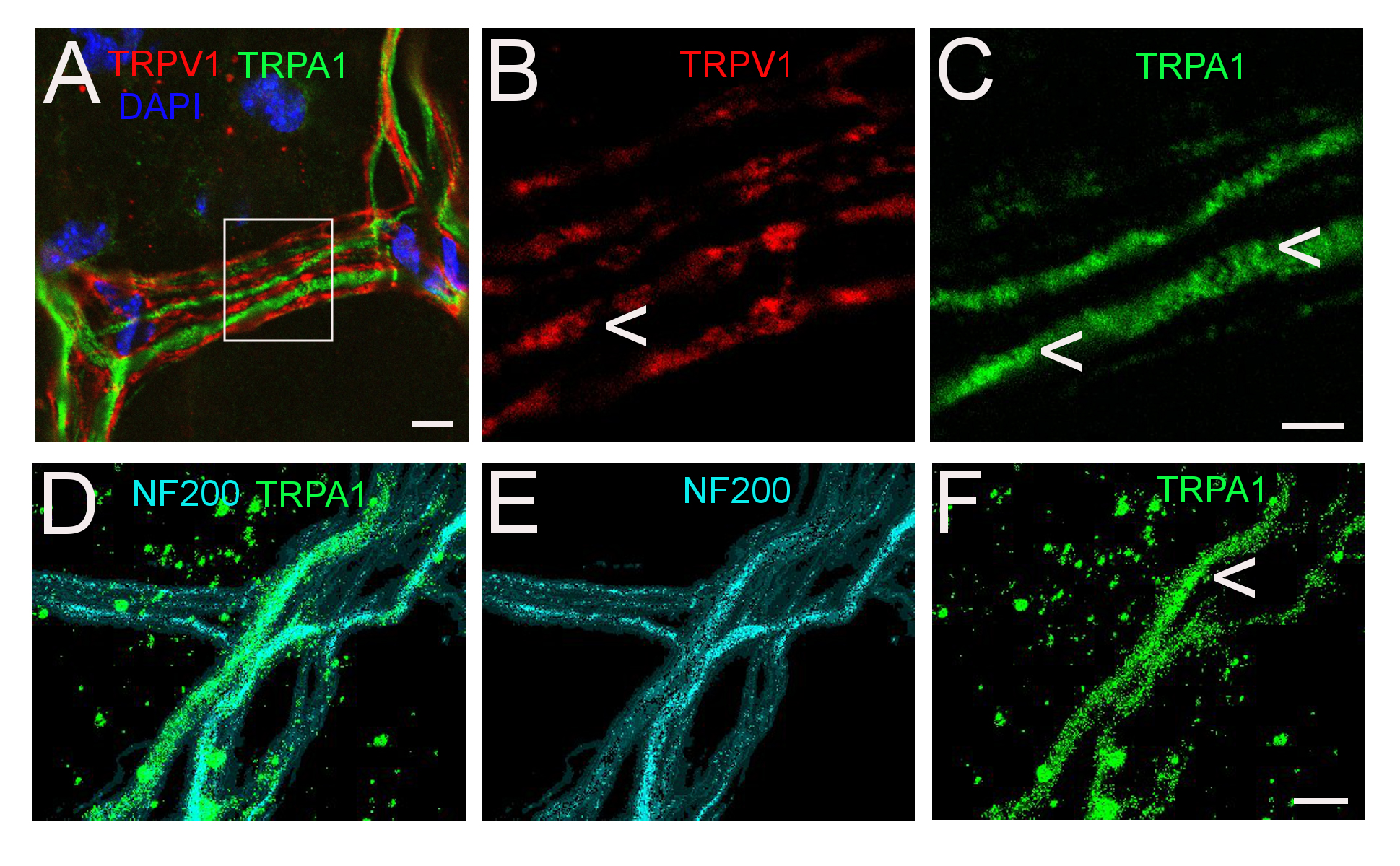
 Figure 2 of
Schecterson, Mol Vis 2020; 26:576-587.
Figure 2 of
Schecterson, Mol Vis 2020; 26:576-587.