Figure 1. TRPV1 is expressed in axons throughout the cornea. A, B: Neuronal β-tubulin (TUJ) and TRPV1 antisera labeled axons in a mouse cornea wholemount. C: Merged section from the wholemount in A, B (boxed area in A) demonstrating many of the axon fiber bundles entering the cornea express TRPV1. D–F: Three-dimensional x,y projection from the sub-basal level to the cornea surface; arrowheads point to small-diameter fibers
expressing TRPV1 extending to the surface. Scale bars: (AB) 500 μm; (C) 350 μm; (D–F) 10 μm.
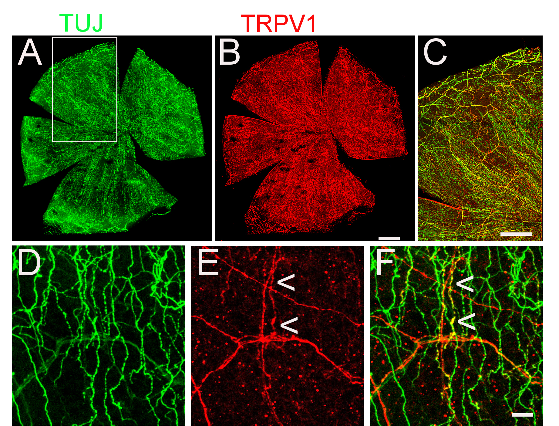
 Figure 1 of
Schecterson, Mol Vis 2020; 26:576-587.
Figure 1 of
Schecterson, Mol Vis 2020; 26:576-587.