Figure 7. IL-6 deficiency significantly prevents PVR progression in vivo. The proliferative vitreoretinopathy (PVR) model was induced in wild-type (WT) and interleukin-6 (IL-6) −/− mice with intravitreal
injection of dispase/collagenase for 7 days. A: Representative hematoxylin and eosin staining images of cross sections of the eyeballs display that the WT mice presented
poorly distinguishable retinal layers, numerous inflammatory cell infiltration (arrow) in the retinal ganglion cell layer
and inner nuclear layer, extensive retinal folds (▲), retinal detachment, and subretinal fibrosis (*). Nevertheless, no obvious
inflammatory cell infiltration and subretinal fibrosis were observed in the retinas of the IL-6 −/− mice. Although localized
retinal folds are still presented, the retinal layers are clearly visible in the IL-6 −/− mice. Retinas were harvested for
quantitative real-time PCR analysis of the expression of IL-6, gp130, and epithelial-mesenchymal transition (EMT) markers.
B: P values compared with the WT group are shown (one-way analysis of variance [ANOVA] with the Tukey-Kramer multiple comparison
test). C: Retinas were harvested for western blotting of the expression of IL-6, gp130, and EMT markers. D: Quantification of IL-6, gp130, and EMT marker protein levels from three independent experiments. P values versus the WT
group are shown (one-way ANOVA with the Tukey-Kramer multiple comparison test).
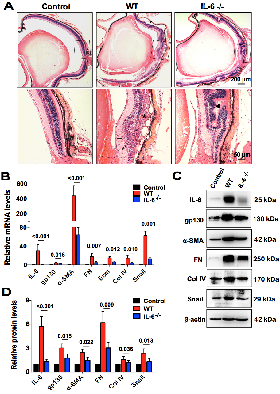
 Figure 7 of
Chen, Mol Vis 2020; 26:517-529.
Figure 7 of
Chen, Mol Vis 2020; 26:517-529.