Figure 5. EMT phenotype is reversed by blocking IL-6/JAK1/STAT3 signaling in RPE cells. A: Microscopic observations of RPE cells exposed to 25 ng/ml of interleukin-6 (IL-6) with or without S3I-201 (5.0 μM) for 48
h. Scale bar: 100 μm. B: Immunofluorescent staining of epithelial-mesenchymal transition (EMT) markers collagen type I, fibronectin, N-cadherin,
and vimentin expression in RPE cells exposed to 25 ng/ml of IL-6 with or without S3I-201(5.0 μM) for 48 h. Scale bars: 40 μm.
C: Western blotting of the expression of EMT markers α-SMA, collagen type I, collagen type IV, fibronectin, N-cadherin, and
Snail in RPE cells exposed to 25 ng/ml of IL-6 with increasing concentrations of S3I-201 (1.25, 2.5, 5.0, and 10.0 μM) for
48 h. D: Quantification of EMT marker protein levels from three independent experiments. P values versus the IL-6 25 ng/ml treatment
group are shown (one-way analysis of variance [ANOVA] with the Tukey-Kramer multiple comparison test).
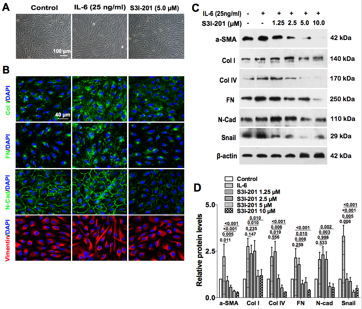
 Figure 5 of
Chen, Mol Vis 2020; 26:517-529.
Figure 5 of
Chen, Mol Vis 2020; 26:517-529.