Figure 1. IL-6 promotes proliferation of RPE cells and EMT. A: Cell counting kit 8 (CCK-8) assay of proliferation of RPE cells after exposure to increasing concentrations of interleukin-6
(IL-6; 5, 10, 25, and 50 ng/ml) for 24, 48, and 72 h. P values versus the control group are shown (one-way analysis of variance
[ANOVA] with the Tukey-Kramer multiple comparison test). B: Microscopic observations of RPE cells in the presence or absence of 10 and 25 ng/ml of IL-6 for 48 h. Scale bar: 100 μm.
C: Western blotting of the expression of epithelial-mesenchymal transition (EMT) markers α-SMA, collagen type I, collagen type
IV, fibronectin, and Snail after treatment with various concentrations of IL-6 (5, 10, 25, and 50 ng/ml) for 48 h. D: Quantification of EMT marker protein levels from three independent experiments. P values versus the control group are shown
(one-way ANOVA with the Tukey-Kramer multiple comparison test).
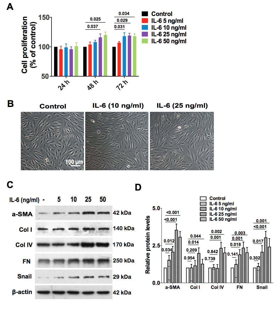
 Figure 1 of
Chen, Mol Vis 2020; 26:517-529.
Figure 1 of
Chen, Mol Vis 2020; 26:517-529.