Figure 2. Immunohistochemical expression of HIF-1α in pterygium and normal conjunctiva. Immunohistochemical expression of HIF-1α in
pterygium (A–B) and normal conjunctiva (C). A: Strong nuclear and cytoplasmic immunoreactivity in all epithelial layers of the pterygium sample. B: A large number of vascular endothelial cells in the stromal layer show brown positive staining. C: Weak cytoplasmic immunoreactivity is detected in the control conjunctival sample (400X, bar = 20 μm).
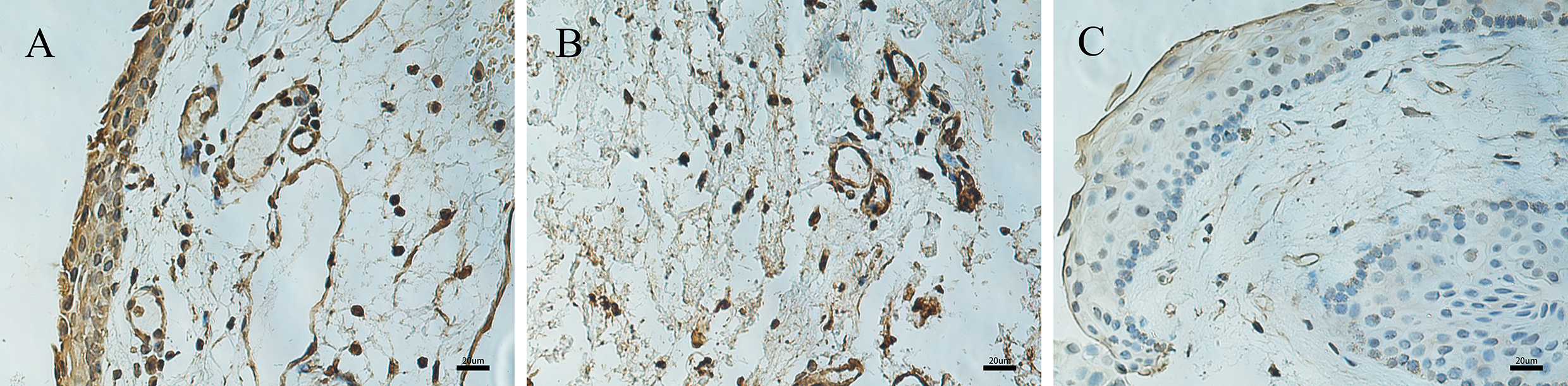
 Figure 2 of
Dong, Mol Vis 2020; 26:510-516.
Figure 2 of
Dong, Mol Vis 2020; 26:510-516.