Figure 4. Analysis of retinal function of mice treated with NaIO3 or NaIO3 + TMP at three time points. A: Rod responses recorded in three groups at day 3, 7, and 14. Statistically significant reductions in the scotopic 0.01 b-wave
amplitudes were observed in the NaIO3-degenerated group (Group I) and the tetramethylpyrazine (TMP) group at three time points. B: Mixed rod-cone responses recorded in three groups at day 3, 7, and 14. The a- and b-wave amplitudes of the mixed rod-cone
responses in the TMP group declined slightly compared with those in Group I at three time points. The a-wave and b-wave latencies
in the TMP group were shorter than those in Group I at day 3 and 7. C: Cone responses recorded in the three groups at day 3, 7, and 14. Significant reductions in the photopic b-wave amplitudes
were observed in Group I compared with those in the TMP group at the day 3 and 7 time points, and significant prolongations
of photopic b-wave latencies were observed in Group I and the TMP group at three time points. (n = 8 retinas from 4 mice,
**p<0.01, *p<0.05).
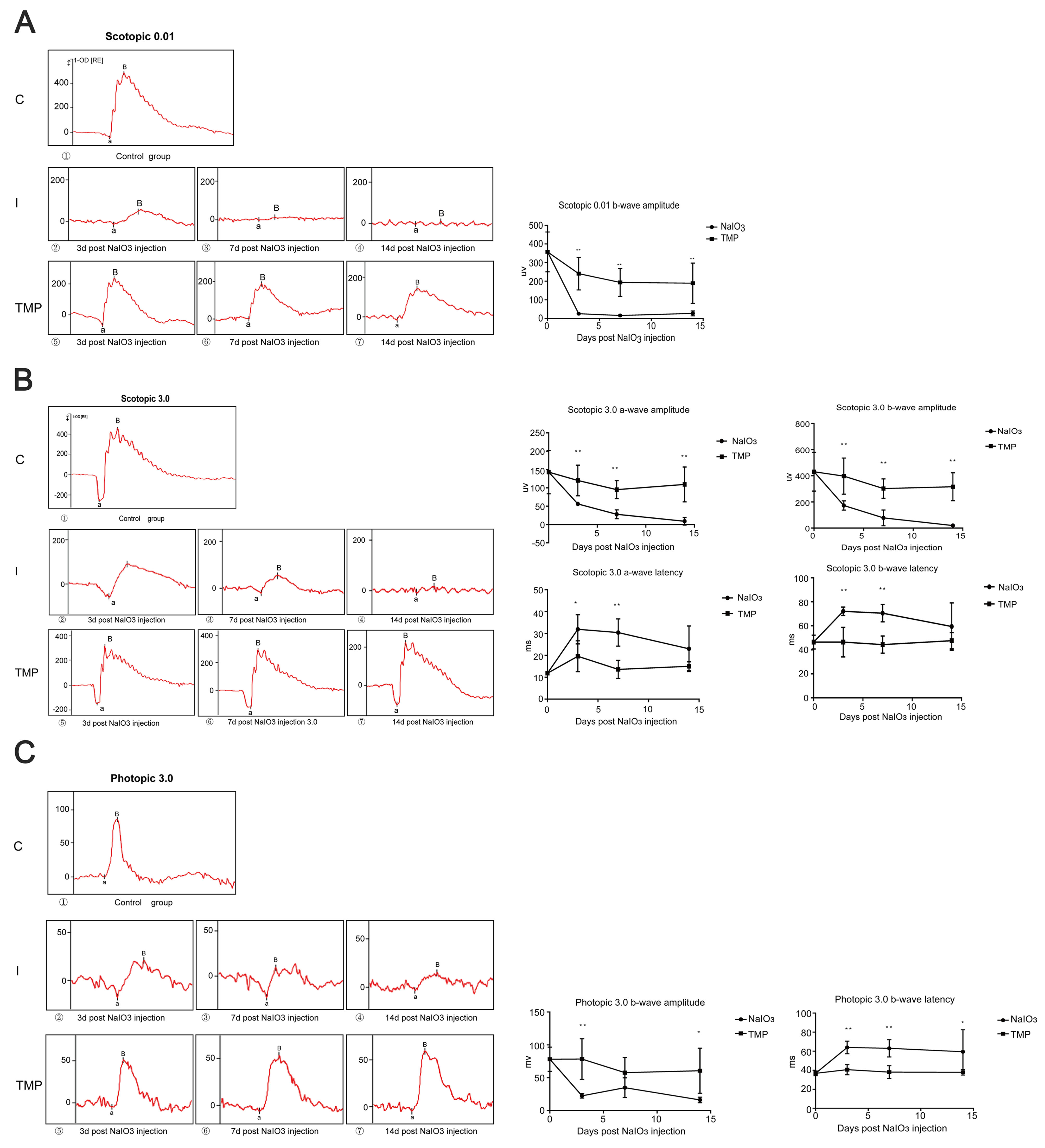
 Figure 4 of
Huang, Mol Vis 2020; 26:494-504.
Figure 4 of
Huang, Mol Vis 2020; 26:494-504.