Figure 2. Images of intravitreal injection into the rat eye. A: Rat eye after administration of atropine sulfate for pupil dilation. B: A string loop was tied around the eye to “bulge out” the eye. C: Betadine iodine was swabbed on the surface of the sclera at the injection site. D: A pilot hole was made in the superotemporal region with a 30-gauge needle on the sclera behind the lens. E: The pilot hole is clearly seen on the sclera. F: A 34-gauge needle attached to a NanoFil syringe was used for injections and was inserted into the pilot hole at a similar
angle. G: The cloudy Invivofectamine 3.0 solution was injected and should be visible through the lens. H: Chlorsig antibiotic was swabbed on the injection site with GenTeal eye gel applied to prevent dryness.
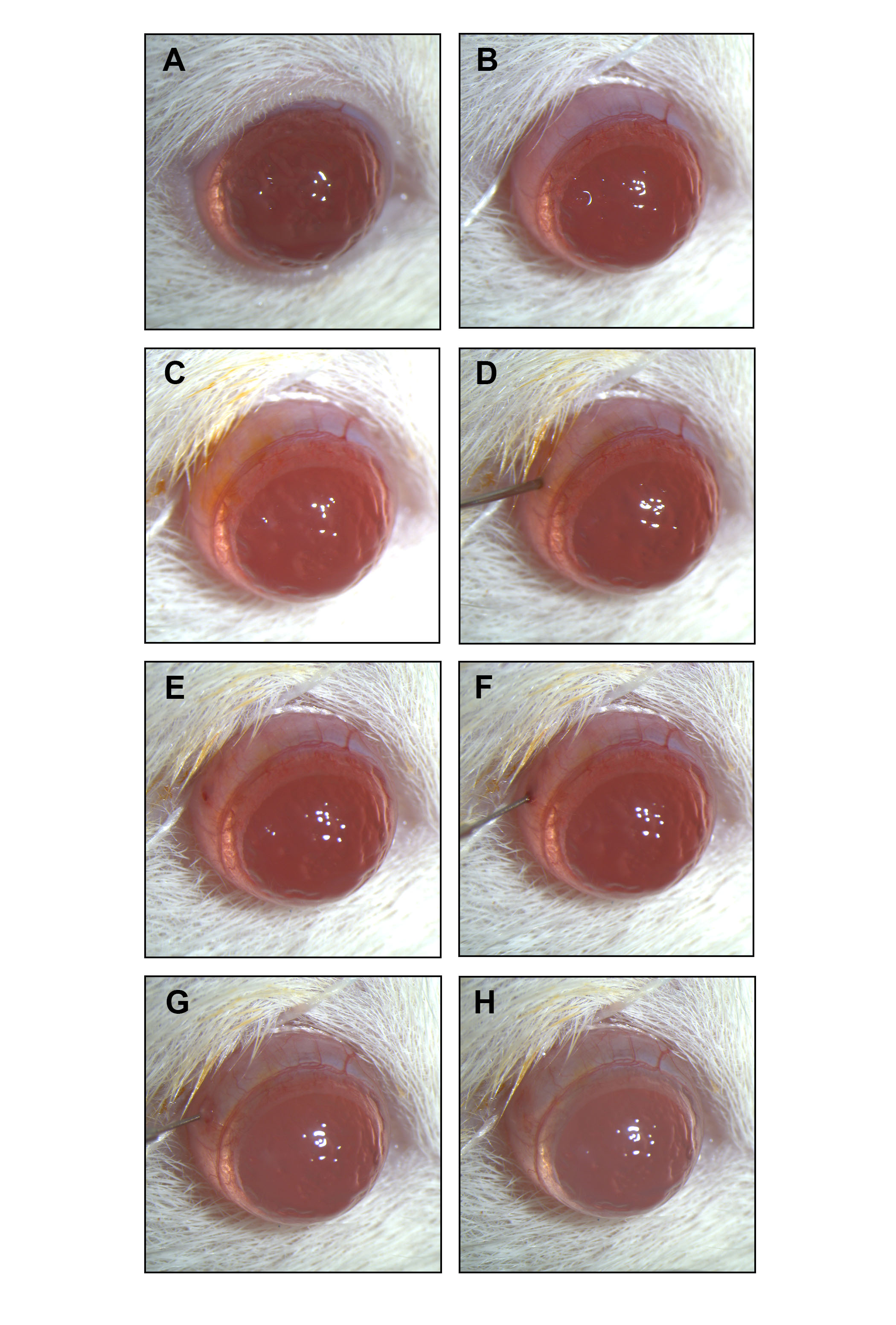
 Figure 2 of
Chu-Tan, Mol Vis 2020; 26:48-62.
Figure 2 of
Chu-Tan, Mol Vis 2020; 26:48-62.