Figure 3. Pathogenic RP1 and RLBP1 variants. The genes are schematically represented and previously reported as retinitis pigmentosa (RP)-causing nonsense or
missense variants marked in RP1. The exons are numbered; white parts indicate non-coding regions, and gray parts indicate coding regions. A: The RP1 gene doublecortin (DCX) domain is marked in green, and the Drosophila melanogaster bifocal (BIF) domain is marked in blue. B: Yellow indicates the retinal-binding domain in RLBP1. The variants of this study are beneath every gene schema with Sanger chromatograms of unaffected heterozygous, and affected
homozygous individuals. Nucleotide variations are circled.
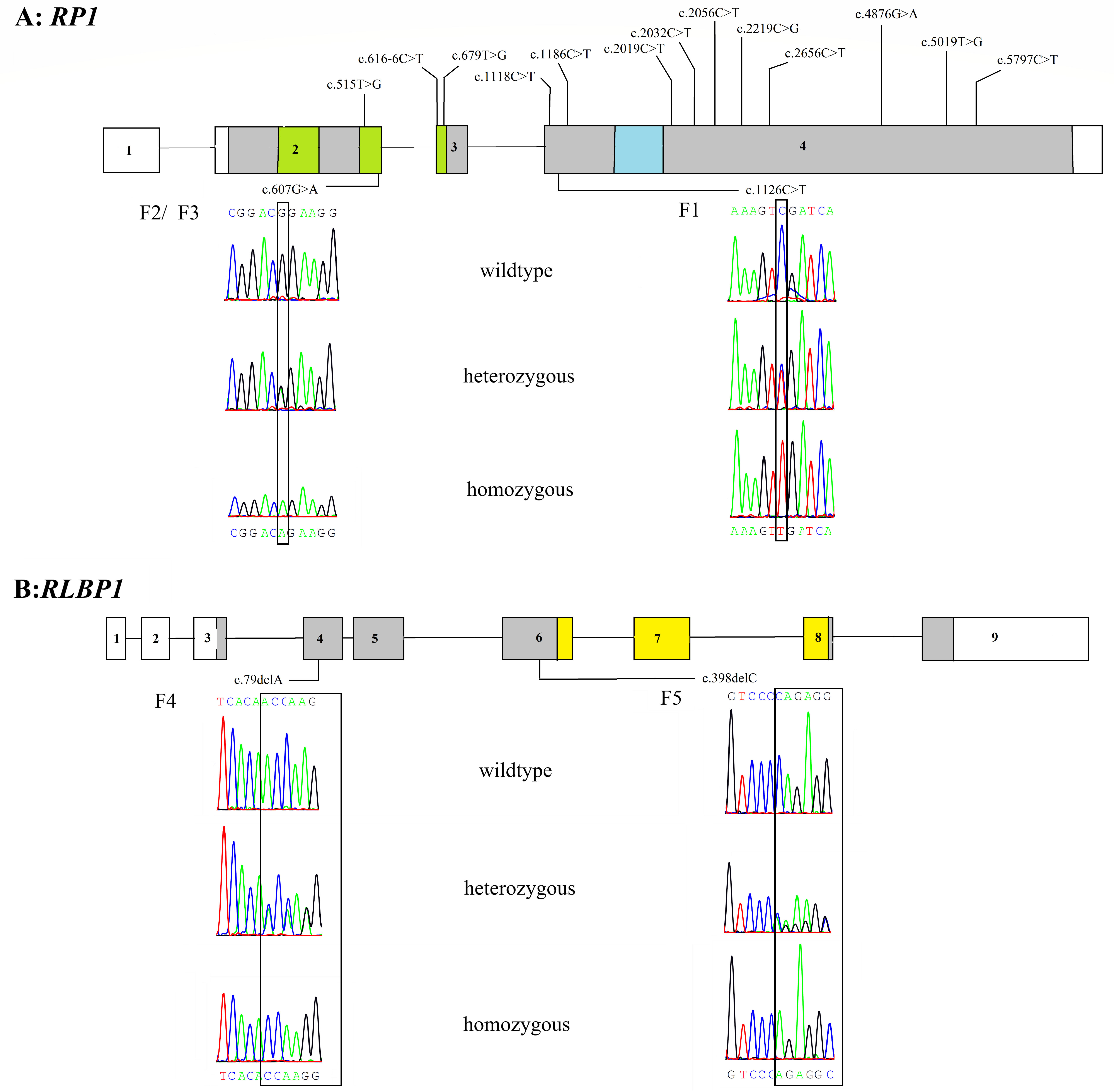
 Figure 3 of
Al-Bdour, Mol Vis 2020; 26:445-458.
Figure 3 of
Al-Bdour, Mol Vis 2020; 26:445-458.