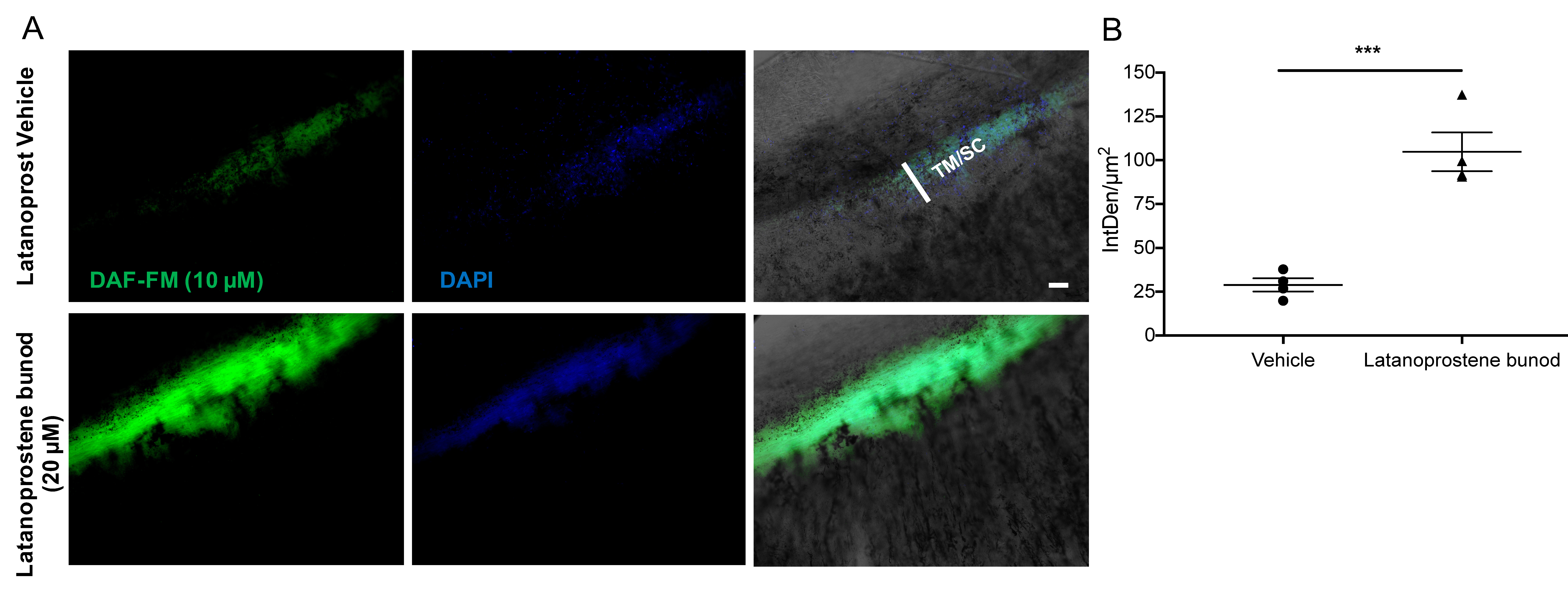
Figure 6. Detection of exogenous NO released from Latanoprostene bunod in human corneoscleral segments using a fluorescent NO-indicator.
A: Increase in DAF-FM fluorescence intensity in quadrants of human corneoscleral segments after treatment with latanoprostene
bunod compared to controls treated with vehicle latanoprost. Quadrants of human donor corneoscleral segments from each eye
(n = 4 per group) were pretreated with intracellular nitric oxide (NO)-indicator dye DAF-FM dye (10 μM) and then treated with
latanoprostene bunod (20 μM) or a latanoprost vehicle. Quadrants treated with latanoprostene bunod showed increased DAF-FM
fluorescence intensity compared with vehicle-treated controls. Images were taken using fluorescence microscopy at 100X magnification
(Scale bar = 50 μm). B: Quantification of DAF-FM fluorescence intensity per unit area (IntDen/μm2) in latanoprostene bunod and latanoprost vehicle treated corneoscleral segments using ImageJ analysis. Data are expressed
as means ± standard error of the mean (SEM); n = 4 for each group; *** p<0.001; two-tailed unpaired Student t test.

 Figure 6 of
Patel, Mol Vis 2020; 26:434-444.
  Figure 6 of
Patel, Mol Vis 2020; 26:434-444.