Figure 3. Detection of nitric oxide (NO) in human corneoscleral segments after treatment with DETA-NONOate/AM (exogenous NO donor).
A: Representative response plot showing amperometric current readings obtained after treatment of ex vivo cultured human corneoscleral
tissues with an exogenous NO-donor DETA-NONOate/AM or equivalent vehicle. A visible spike in recorded current was observed
as a result of DETA-NONOate/AM treatment on human corneoscleral segments. A “no tissue” control was employed to ensure that
the detection of NO signal was tissue dependent and not a result of NO release in aqueous PBS. B: Change in NO concentration from baseline after 30 min of treatment with DETA-NONOate/AM (20 μM) or equivalent vehicle (0.1%
dimethyl sulfoxide [DMSO]) at room temperature. Data are expressed as means ± standard error of the mean (SEM); n = 5 for
each group; ** p<0.001; two-tailed unpaired Student t test.
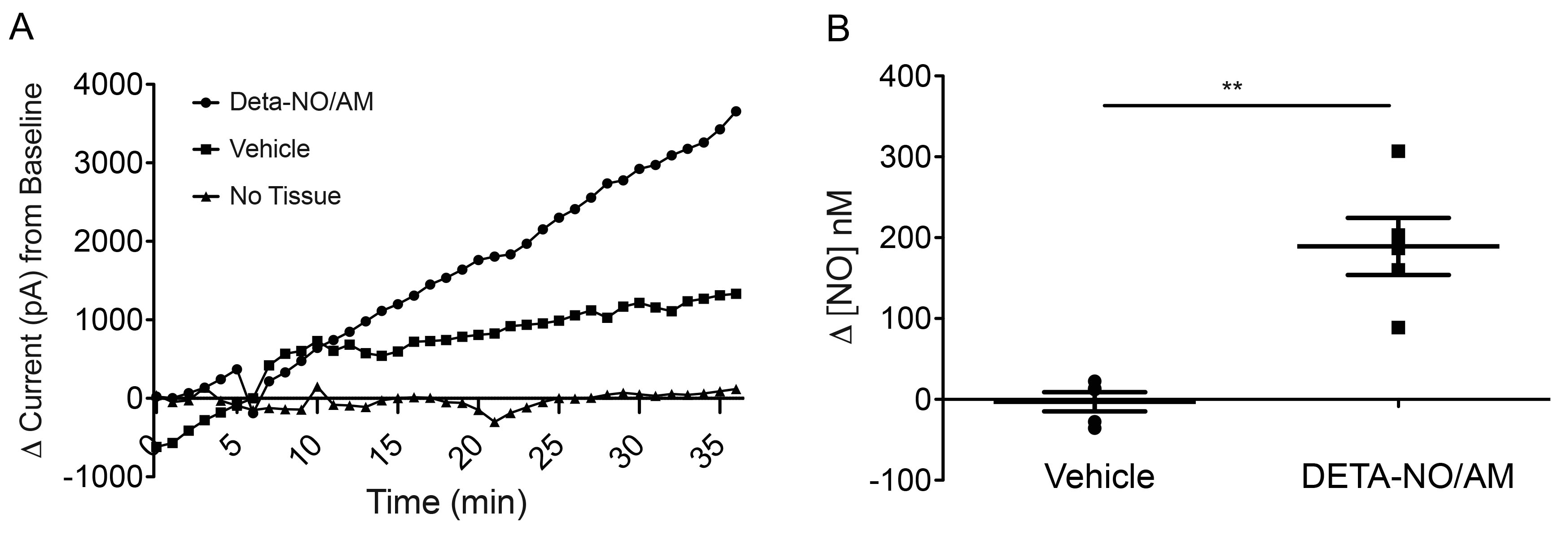
 Figure 3 of
Patel, Mol Vis 2020; 26:434-444.
Figure 3 of
Patel, Mol Vis 2020; 26:434-444.