Figure 1. Experimental setup for the study. A: Workflow for electrochemical nitric oxide (NO) measurement using NO-sensing electrode. Cultured human donor corneoscleral
segments (C = cornea; S = sclera; TM = trabecular meshwork) were washed 3X with PBS and placed in a 24-well plate with 1 ml
of PBS. Drug treatments were performed after a stable baseline was achieved. The output current corresponds to the level of
NO generated (~10 pA/nM), detected using a free radical analyzer (TBR1025). The signal is amplified by a four-channel Lab-Trax
amplifier and analyzed using LabScribe3 software. B: Workflow for fluorochemical measurement of NO using DAF-FM assay (intracellular NO-binding dye). Quadrants of the human donor
corneoscleral segment were cultured at 37 °C in DMEM media supplemented with 2% fetal bovine serum (FBS) and 1% penicillin–streptomycin
(PS) and treated with 10 μM NO-binding fluorescent DAF-FM dye for 30 min. Cells and tissues were washed 1X with PBS and treated
with different drugs or vehicle and incubated at 37 °C for an additional 30 min. Tissues were then washed 3X with PBS, and
the trabecular meshwork (TM) rim was removed and imaged using fluorescence microscopy.
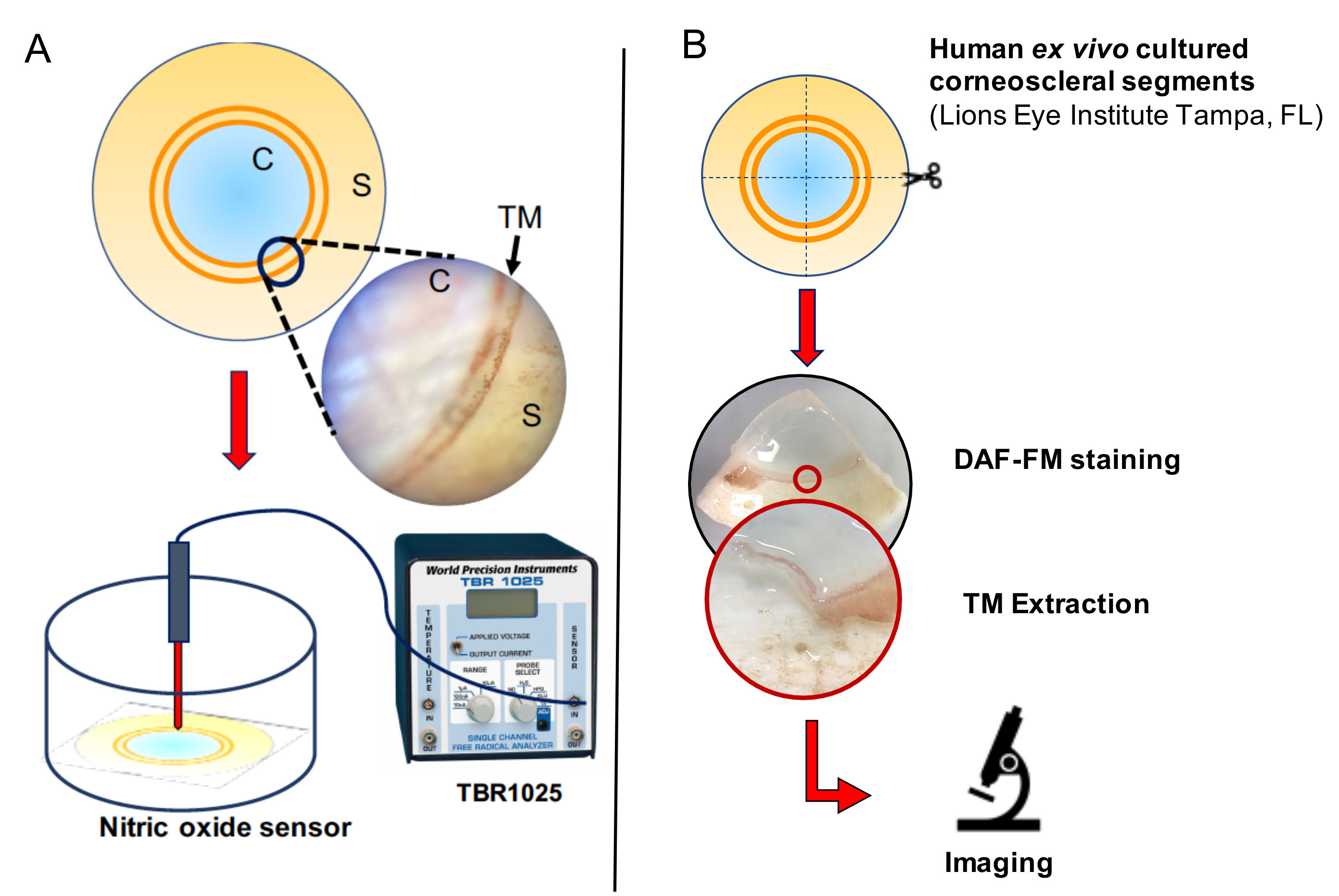
 Figure 1 of
Patel, Mol Vis 2020; 26:434-444.
Figure 1 of
Patel, Mol Vis 2020; 26:434-444.