Figure 2. Retinal imaging from patients with a representative spectrum of biallelic RP1-associated disease. Imaging for each patient includes fundus photography (top image), fundus autofluorescence (middle image),
and OCT (bottom image). The patients shown had macular dystrophy (A: MEE1, images acquired at age 38), cone-rod dystrophy (B: CEI26529, images acquired at age 15), adult-onset retinitis pigmentosa (C: MEE5, images acquired at age 46), and early-onset RP (D: MEE9, images acquired between ages 45–47).
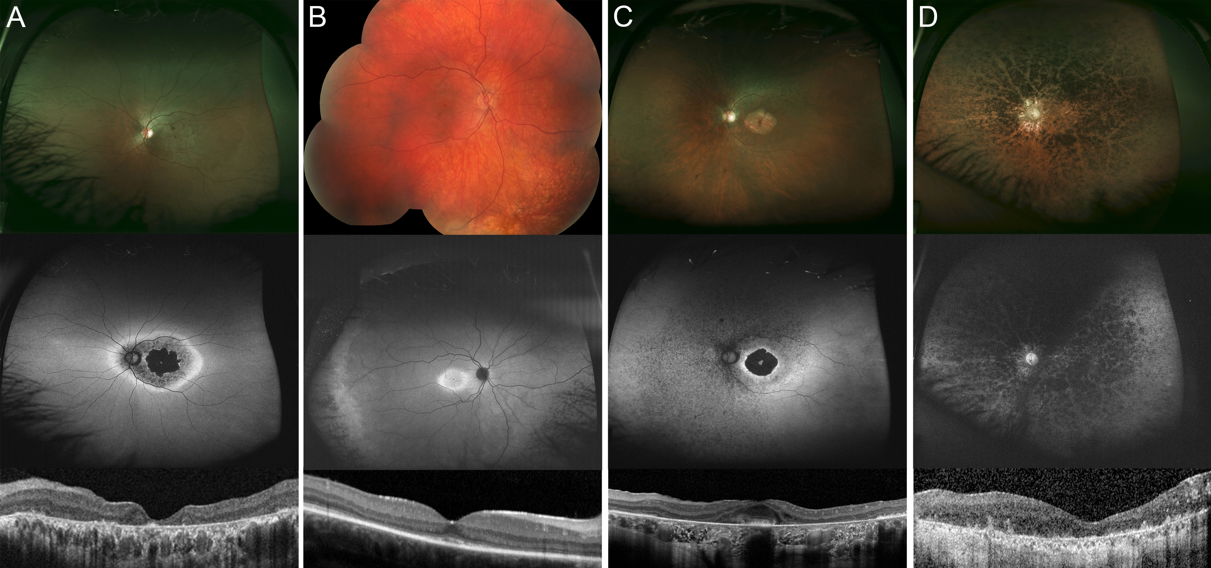
 Figure 2 of
Huckfeldt, Mol Vis 2020; 26:423-433.
Figure 2 of
Huckfeldt, Mol Vis 2020; 26:423-433.