Figure 2. Diagrammatic representation of the retinal flatmount showing areas of RGC count. Three microscopic fields (120 x 160 µm2) in each quadrant were chosen to count labelled RGCs. Area labelled “C” was 0.875 mm from the optic disc. The distance between
areas A, B, and C was 1 mm.
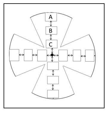
 Figure 2 of
Jafri, Mol Vis 2020; 26:392-408.
Figure 2 of
Jafri, Mol Vis 2020; 26:392-408.