Figure 4. Photo of the slit-lamp microscope and IVCM. A and B: Photo of the slit-lamp microscope and IVCM. There was no significant difference in CNFD among the groups (p=0.2116) or in
the G-DESOS group on Days 0 and 30 (p=0.1926). More dendritic cells clustered in the corneal epithelial layer in the C-DESOS
and G-DESOS groups compared to the CMDE and control groups (p=0.0052). After glucocorticoid treatment, the number of dendritic
cells decreased significantly (p<0.001).
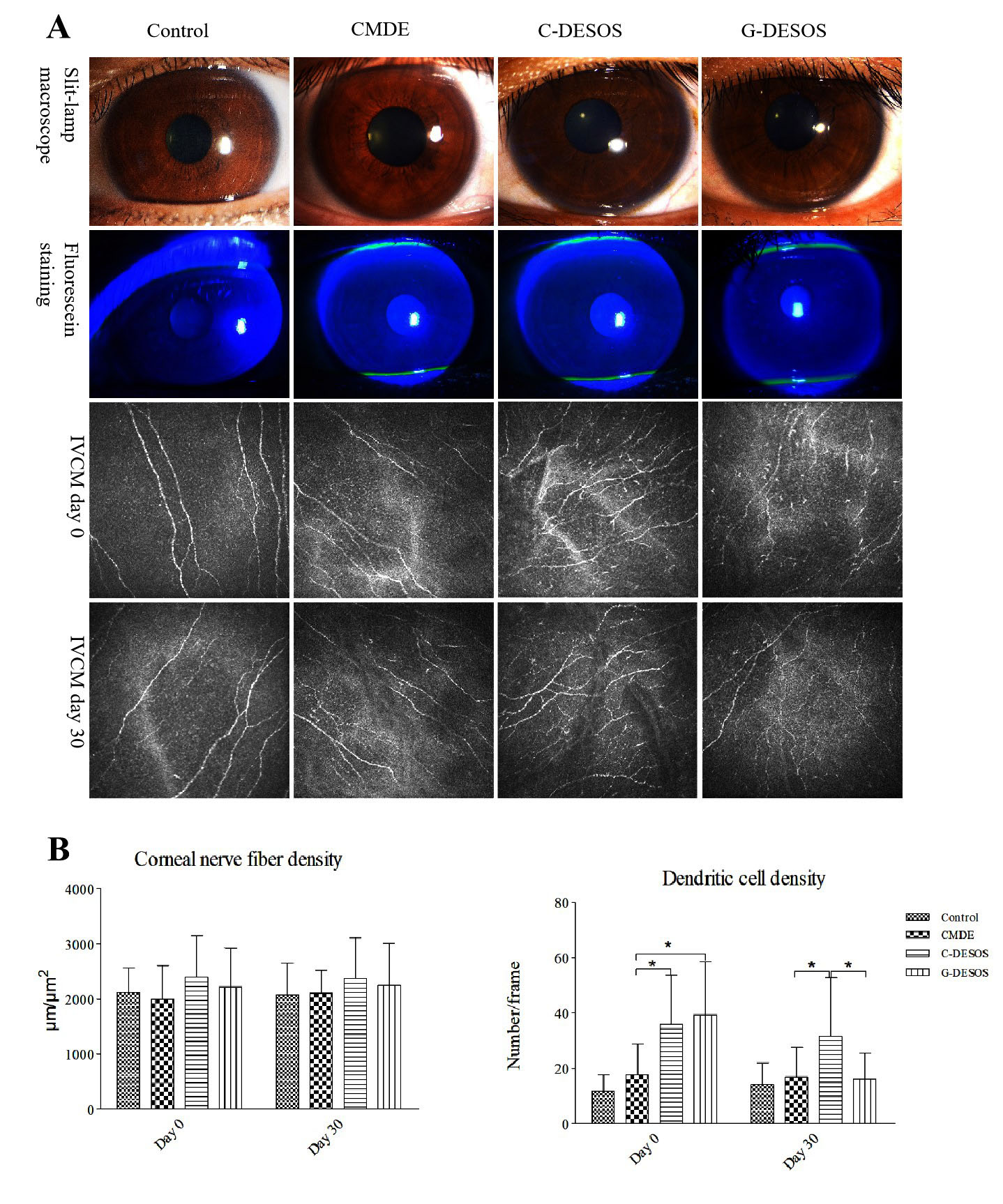
 Figure 4 of
Li, Mol Vis 2020; 26:359-369.
Figure 4 of
Li, Mol Vis 2020; 26:359-369.