Figure 5. Histopathological findings in skin from FAF II-5 subject. A:Histological sections from the eyelid skin biopsy, stained with hematoxylin and eosin and Congo red, showing thin skin with
mild orthokeratotic hyperkeratosis, papillomatosis, mild inflammatory infiltrate composed of perivascular lymphocytes in the
superficial dermis and focally pigmentary incontinence. Dermal appendices appear normal. Congo red staining indicates amyloid
deposits composed of internal degradation fragments produced during the aberrant processing in the hair follicles (arrows).
B: Eyelid skin biopsy shows fibroconnective tissue, skeletal muscle, and vessels (arrows). The Congo red staining is positive
in the walls of the vessels.
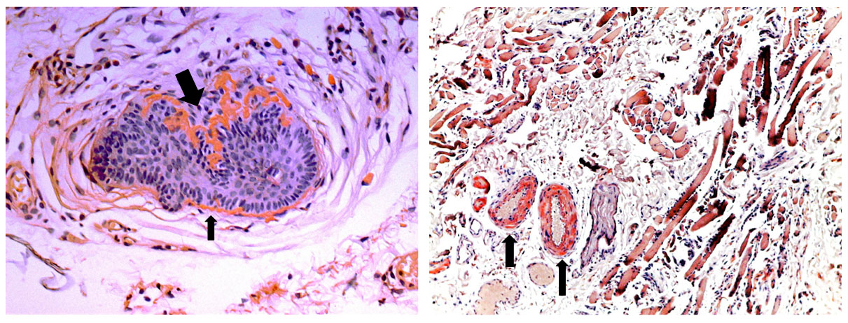
 Figure 5 of
Cabral-Macias, Mol Vis 2020; 26:345-354.
Figure 5 of
Cabral-Macias, Mol Vis 2020; 26:345-354.