Figure 3. Slit-lamp photographs of affected subjects II-2, II-5, and II-6. Multiple central and peripheral lattice-pattern lines and
thin dots with centripetal distribution are evident at the subepithelium and anterior corneal stroma. On retroillumination,
branching refractile lines were evident in all three patients. A: Right eye. B: Left eye.
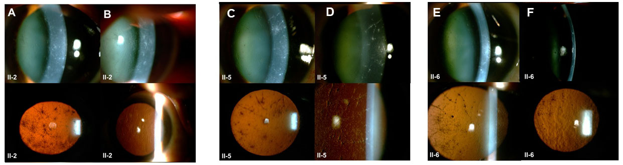
 Figure 3 of
Cabral-Macias, Mol Vis 2020; 26:345-354.
Figure 3 of
Cabral-Macias, Mol Vis 2020; 26:345-354.