Figure 3. Retinal imaging by fundoscopy of affected members. Representative color fundus photos of five affected individuals are shown.
Individual numbers and the age in years are shown on the left. MOL1154 III:1, II:4, and II:2 - confluent foveal drusen-like
clusters, while no significant findings are evident in the periphery. MOL1154 III:3 - advanced macular atrophy in both eyes
with pigmentary dots and patches in RE and a yolk-like lesion surrounded by atrophic retina temporal to the fovea in LE. MOL1154
IV:2 - refractile drusen-like dots surrounding macular atrophy containing yellowish deposits. OD (oculus dextrus) - right
eye; OS (oculus sinister) - left eye.
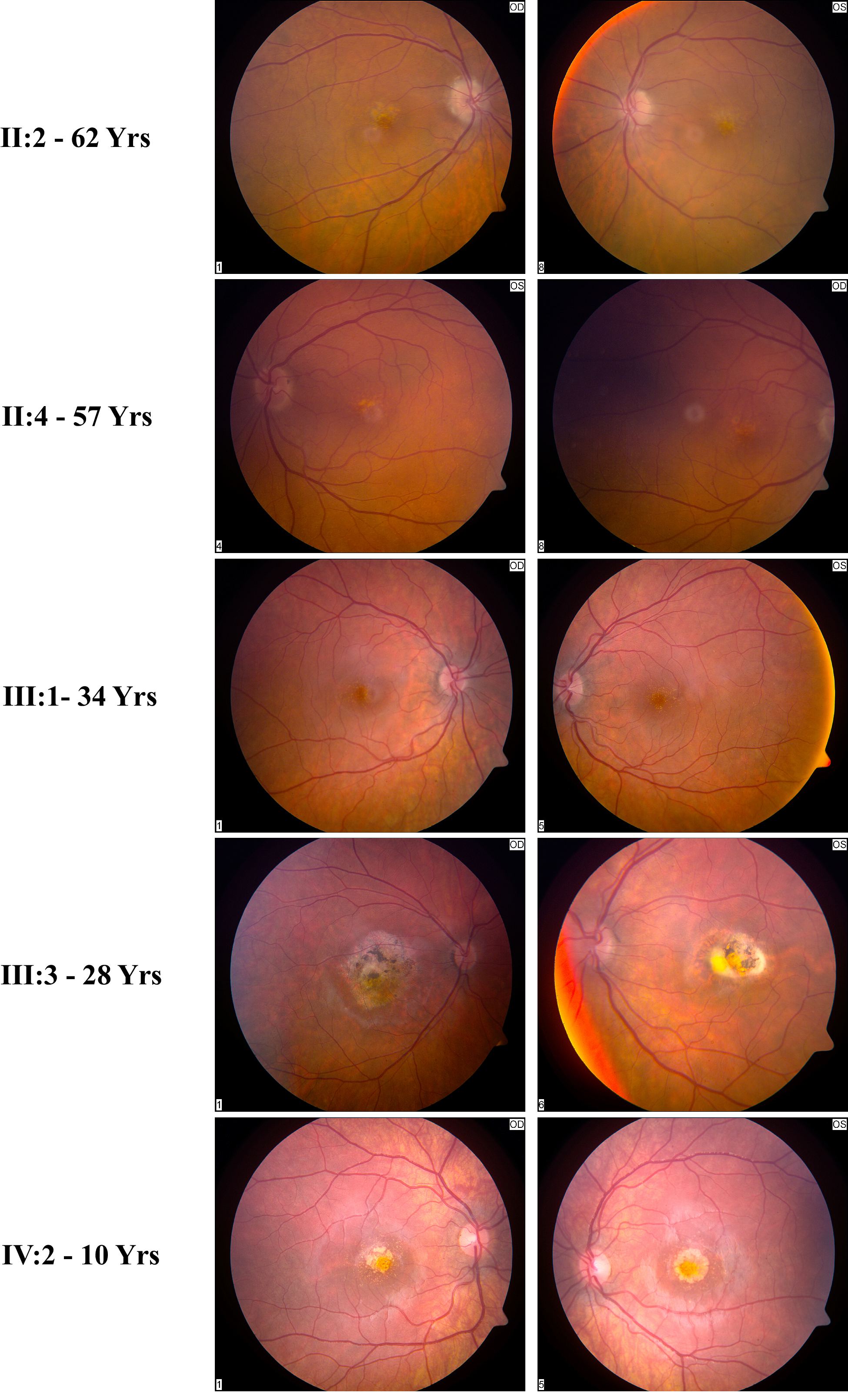
 Figure 3 of
Namburi, Mol Vis 2020; 26:299-310.
Figure 3 of
Namburi, Mol Vis 2020; 26:299-310.