Figure 2. OCT images of affected family members. Representative OCT scans of five affected individuals are shown. Individual numbers
and the age in years are shown on the left. MOL1154 I:2 - preserved inner retinal layer combined with granular interdigitation
layer. MOL1154 II:2 and II:4 - pseudodrusens with intact outer and inner retinal layers. MOL1154 III:1- OCT cross-sections
show normal retinal structure. Note that drusen-like deposits were seen clinically but do not appear in the OCT sections.
MOL1154 IV:2 - subfoveal RPE atrophy combined with thickening of the interdigitation layer, while the ellipsoid layer seems
to be intact.
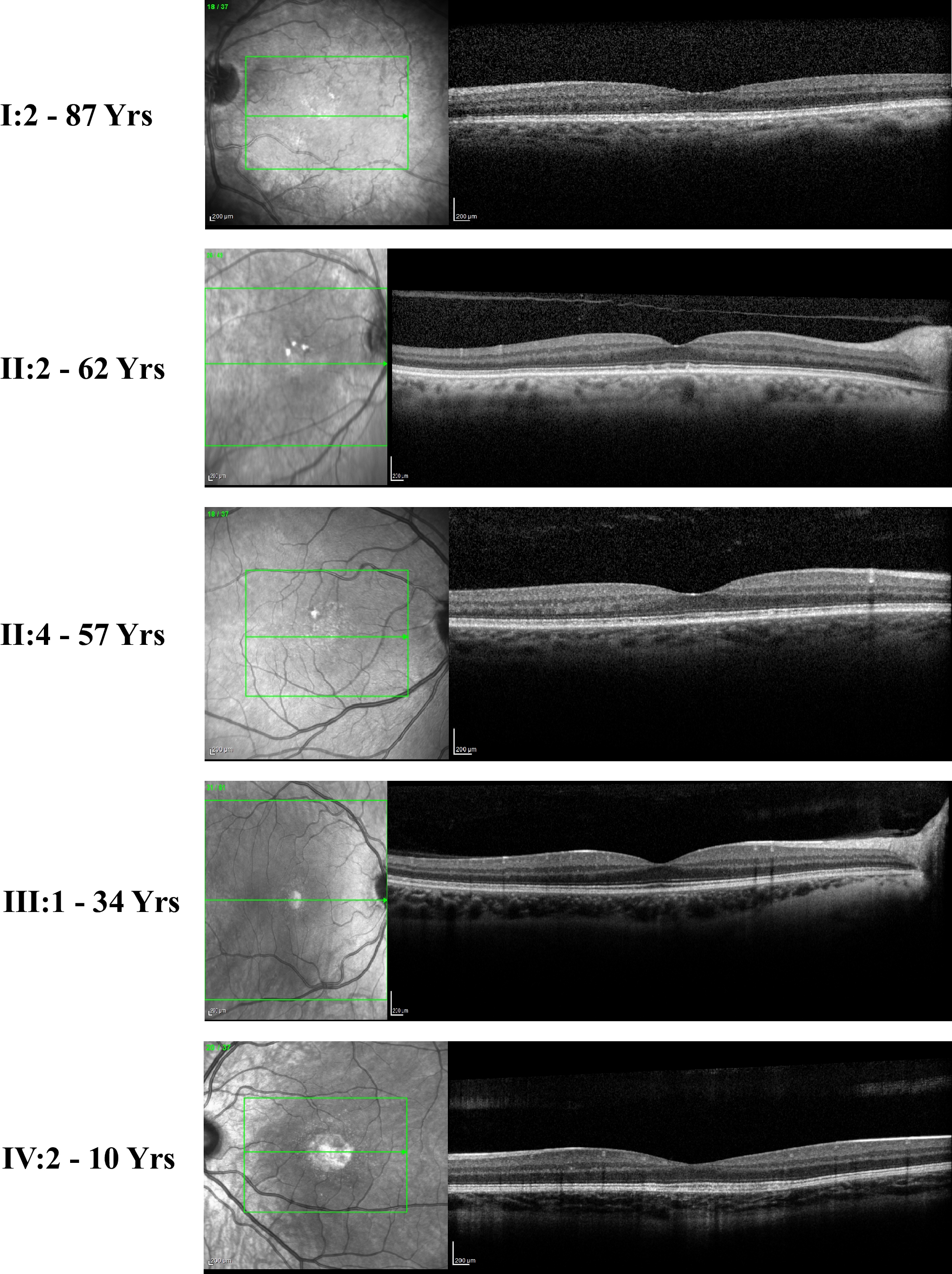
 Figure 2 of
Namburi, Mol Vis 2020; 26:299-310.
Figure 2 of
Namburi, Mol Vis 2020; 26:299-310.