Figure 3. Colored fundus (CF) photographs and optical coherence tomography (OCT) scans of four patients with X-linked retinoschisis
displaying atypical fundus appearances. A: CF photographs of patient 113270 showing bilateral peripheral retinoschisis involving the macula and severe retinal pigment
degeneration with pigmentation, sheathed retinal vessels, and white dots at the temporal retina. B: CF photographs of patient 019143 presenting bilateral macular atrophy and retinal pigment degeneration with bone spicules
and arteriole narrowing in the peripheral retina. C: CF photographs of patient 113580 showing macular atrophy with pigmentation, white spiculations in the peripheral retina
of the right eye, and pigmentation in the peripheral retina of the both eyes and OCT scans presenting bilateral macular atrophy
and peripheral retinoschisis in the right eye. D: CF photographs and OCT scans of patient 1131090 displaying symmetric macular atrophy with pigmentation.
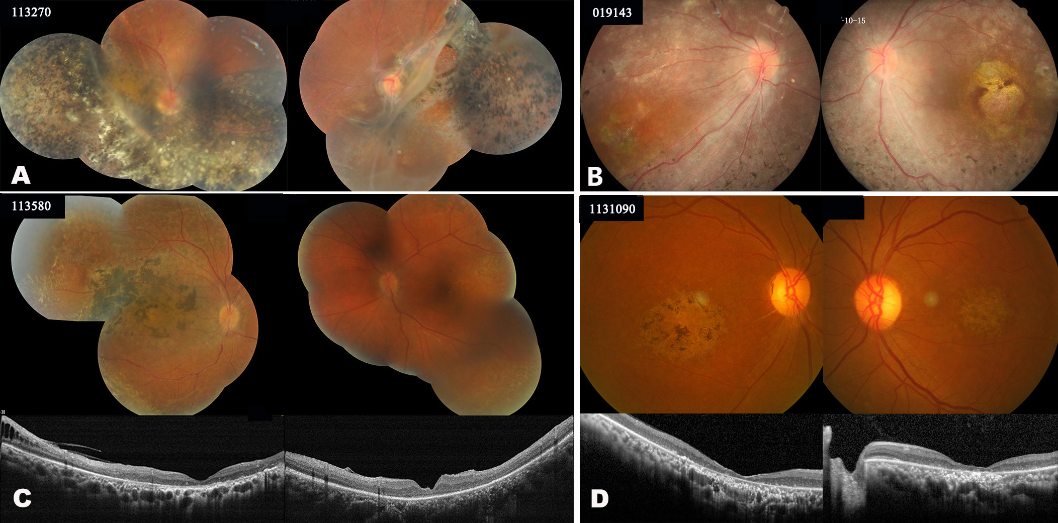
 Figure 3 of
Chen, Mol Vis 2020; 26:291-298.
Figure 3 of
Chen, Mol Vis 2020; 26:291-298.