Figure 2. Colored fundus (CF) photographs, optical coherence tomography (OCT) scans, and fundus autofluorescence (FAF) of five patients
with RS displaying typical macular and peripheral retinoschisis or macular atrophy. A: CF photographs, OCT scans, and FAF of patient 113690 showing macular retinoschisis and a spoke-wheel pattern of hyperfluorescence.
B: CF photograph and OCT scan of patient 113190 displaying macular retinoschisis. C: CF photograph, OCT scan, and FAF of patient 113370 displaying macular atrophy and hypofluorescence in the macular region.
D: CF photograph and OCT scan of patient 113660 presenting both macular and peripheral retinoschisis. E–H: CF photograph and OCT scan of patient 113310 showing normal macular structure (E and F) and peripheral retinoschisis (E and G). H: FAF of patient 113310 presenting almost-normal fluorescent pattern in the macular region and a hypofluorescent region corresponding
to the peripheral retinoschisis.
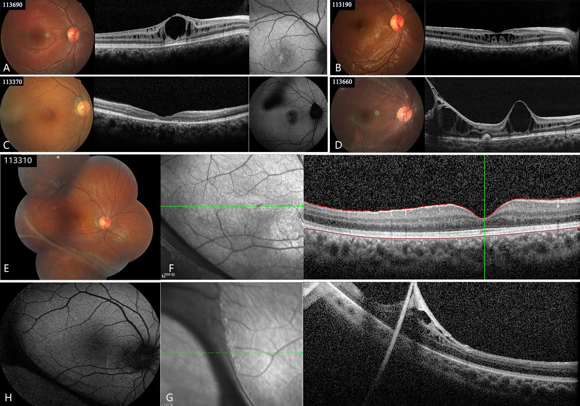
 Figure 2 of
Chen, Mol Vis 2020; 26:291-298.
Figure 2 of
Chen, Mol Vis 2020; 26:291-298.