Figure 5. Representative images of immunogold labeling in retinal explants. The retinal explants (n = 4 for each group) were incubated
in physiological buffer, high glucose alone and in combination with proinflammatory cytokines followed by immunolabeling for
glutamate (A–C), glutamine (D–F), GABA (G–I), and glycine (J–L). The retinal layers of interest are indicated on the left side of panel A. The two horizontal lines on the left side of each image identify the IPL. Cells of interest are indicated in A with the bipolar cell, identified in the INL close to the OPL (BC, red arrow), the stellar-shape soma of Müller cells in
the middle of the INL (MC, blue arrow), the amacrine cells in the lower row of the INL adjacent to the IPL (AC, yellow arrow),
the cells in the ganglion cell layer (GC, orange arrow), and the intercellular space corresponding to Müller cell endfeet
in the GCL (MCef, black arrowhead). A: Glutamate labeling in the control spans the entire retina except the middle row of nuclei in the INL, which corresponds
to Müller cells. However, MCef displayed increased glutamate labeling in high glucose alone (B, arrowhead and insert) and in combination with proinflammatory cytokines (C, arrowhead and insert). Coapplication of high glucose and proinflammatory cytokines also appears to decrease glutamate labeling
in bipolar cells (C, red arrows). D: Glutamine labeling in the control retina was seen predominantly in the inner retinal layers. A decrease
in glutamine distribution was apparent in the MCef in the high glucose and proinflammatory cytokine condition (F, arrowhead) but not in high glucose alone (E). GABA labeling in the control retina was evident in amacrine cells, IPL, and displaced amacrine cells in the GCL (G), with no apparent differences in high glucose alone (H) or in combination with proinflammatory cytokines (I). Glycine immunolabeling was seen in amacrine cells in the control retina (J) with no apparent differences in high glucose alone (K) or in combination with proinflammatory cytokines (L). Scale bar is equal to 25 µm. Abbreviations: AC, amacrine cell; BC, bipolar cell; GC, ganglion cell; GCL, ganglion cell
layer; INL, inner nuclear layer; IPL, inner plexiform layer; IS, photoreceptor inner segment; MC, Müller cell; MCef, Müller
cell endfeet; ONL, outer nuclear layer; OPL, outer plexiform layer; OS, photoreceptor outer segment.
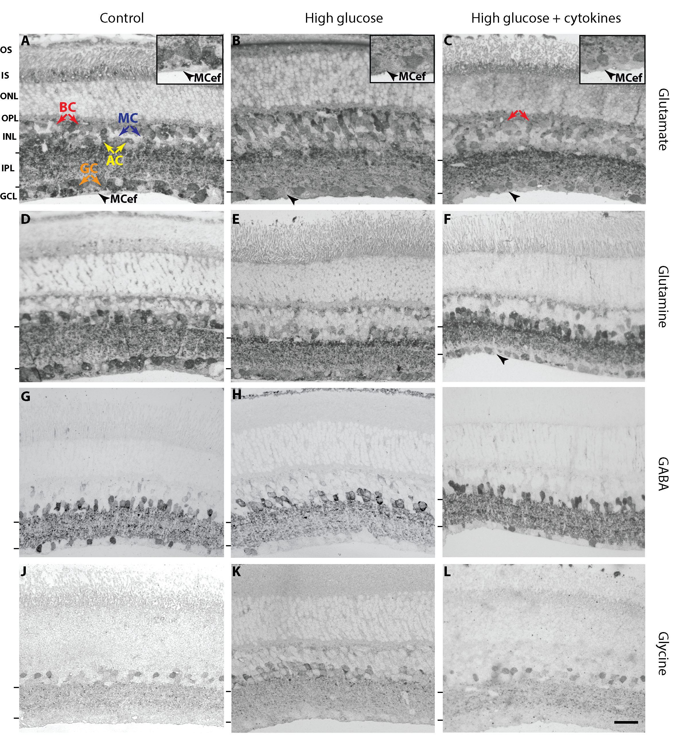
 Figure 5 of
Shivashankar, Mol Vis 2020; 26:277-290.
Figure 5 of
Shivashankar, Mol Vis 2020; 26:277-290.