Figure 2. Two patients with nephronophthisis and Caroli disease with homozygous mutations in WDR19 c.3533G>A. A–D: Fundus photograph, optical coherence tomography, renal ultrasonography, and abdominal computed tomography of a 7-year-old
patient with Leber congenital amaurosis (LCA) with a homozygous mutation in WDR19 c.3533G>A (P36). Increased kidney echogenicity, multiple cystic formation of the kidney, and dilated intrahepatic bile duct
were noted. She was neurologically normal. E–H: Fundus photograph, renal and abdominal ultrasonography, and brain magnetic resonance imaging (MRI) image of a 4-year-old
patient with possible dual diagnosis of mutations in WDR19/POLG (P37). Brain MRI showed a bilateral T2 hyperintense signal in the putamen, which was not reported in Senior-Løken syndrome
(black arrow). These findings suggest a possible dual diagnosis.
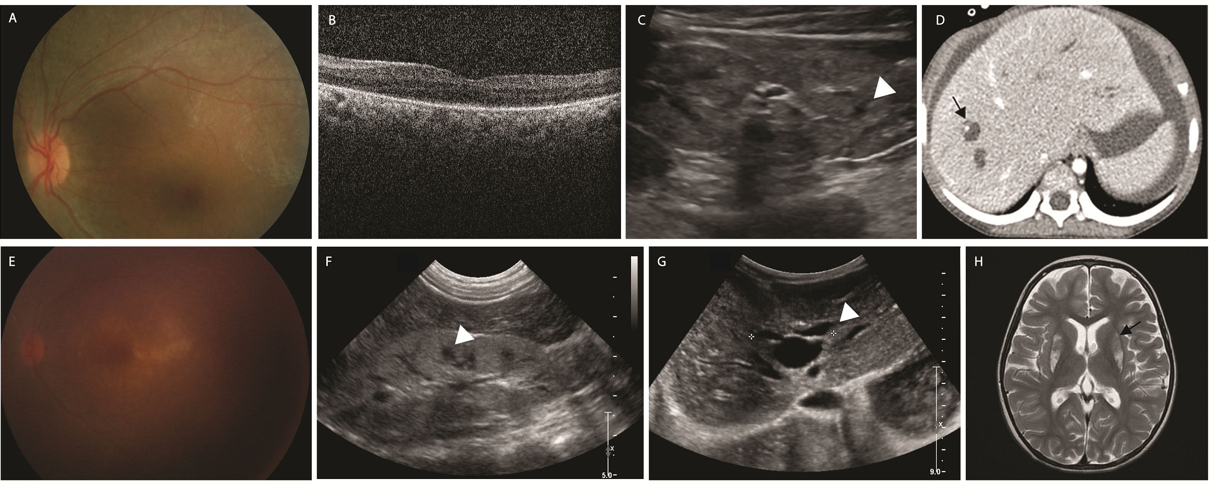
 Figure 2 of
Surl, Mol Vis 2020; 26:26-35.
Figure 2 of
Surl, Mol Vis 2020; 26:26-35.