Figure 3. Synaptic morphology at P19, P24, P32, and P47 in RA and OIR mice. A and B: Immunohistochemistry showing ectopically aligned synapses between photoreceptor presynaptic terminals and rod bipolar cells
in the OPL from retinal cross sections at P19, P24, P32, and P47. PKC-alpha (red in A, white in B) stains rod bipolar cells, and PSD95 (green in A) stains photoreceptor presynaptic terminals. C: Quantification of ectopic dendrites per 200 µm in the central and peripheral retina. The white arrow heads in A and B point to ectopically localized synapses extending beyond the OPL. RA = room air; OIR = oxygen-induced ischemic retinopathy;
P = postnatal day; OPL = outer plexiform layer; PSDS = postsynaptic density protein; PKC-alpha denotes protein kinase c alpha.
Three RA mice and three OIR mice were examined for each developmental age studied. Stars denote level of significance: *p
< 0.05, **p < 0.01, ***p < 0.001, and ****p < 0.0001. Sampling size: n=3 for each group. Error bars represent SEM.
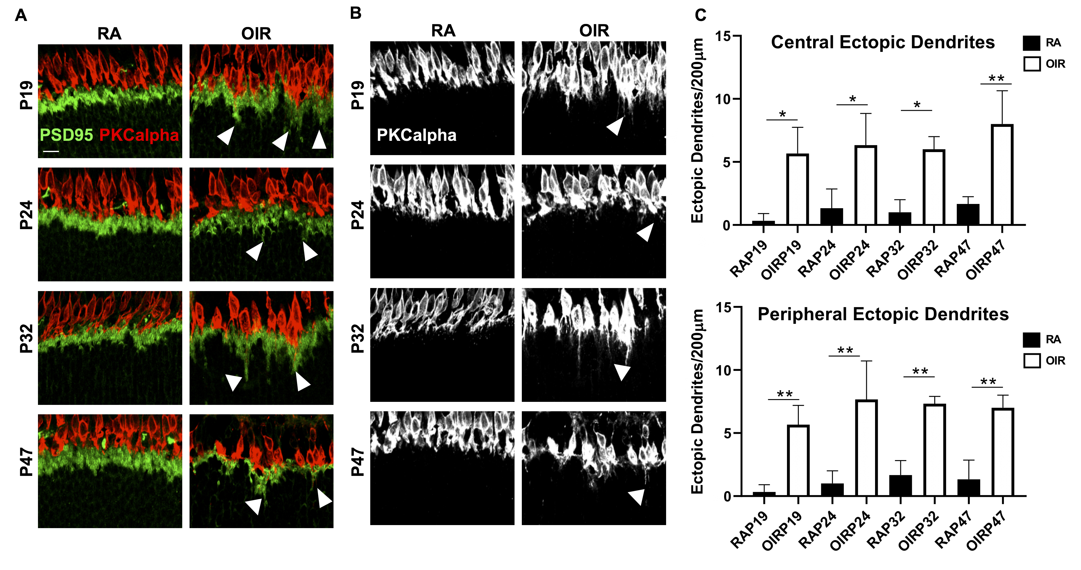
 Figure 3 of
Mezu-Ndubuisi, Mol Vis 2020; 26:257-276.
Figure 3 of
Mezu-Ndubuisi, Mol Vis 2020; 26:257-276.