Figure 3. Coimmunostaining of P2 foveal glial cells with the Müller cell marker, GS, GFAP, and vimentin. Coimmunostaining of foveal
cryosections from P2 with glutamine synthetase (GS), which is a specific marker of Müller cells in humans, glial fibrillar
acidic protein (GFAP), and vimentin (VIM), which stains all glial origin cells, shows that the GFAP-positive cells at the
roof of the foveal pit (A, B, and inset) are stained with vimentin (C, D, and inset) but do not express GS (E, F, and inset). Note that vimentin and GS stain the retinal Müller glial cells of the fovea (C, white arrow and inset black arrow) and the Z-shaped Müller cells of Henle’s fiber layer (D). GCL, ganglion cell layer; INL, inner nuclear layer; ONL, outer nuclear layer; HL, Henle’s fiber layer; OLM, outer limiting
membrane.
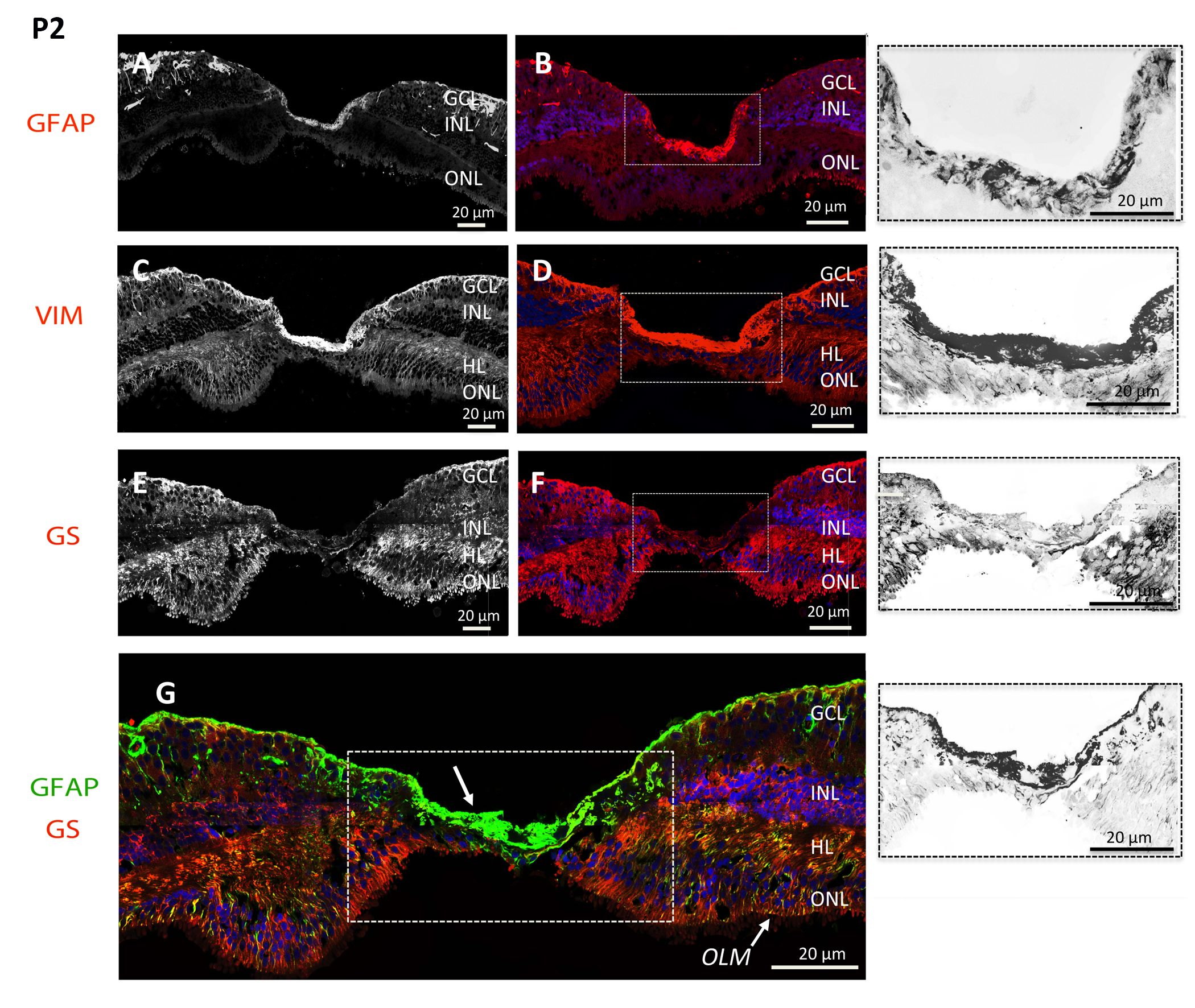
 Figure 3 of
Delaunay, Mol Vis 2020; 26:235-245.
Figure 3 of
Delaunay, Mol Vis 2020; 26:235-245.