Figure 1. Cryosections and immunolabeling of cells at the roof of the foveal pit. Cryosections (10 µm thick) of the foveola from P1
(A), P2 (B), and P4 (C) showing the presence of cell nuclei (4′, 6-diamino-2-phenylindole [DAPI] staining) at the roof of the foveal pit (arrows).
Costaining with the glial marker glial fibrillar acidic protein (GFAP) of the foveola from P1 (D), P2 (E), and P4 (F) shows that GFAP-positive cells with lateral extension lie on the roof of the fovea (black and white higher magnification
insets). The nuclei of those cells are in the innermost layer and do not send radial extensions toward the outer nuclear layers,
demonstrating that these cells are distinct from Müller cone cells. GCL, ganglion cell layer; INL, inner nuclear layer; ONL,
outer nuclear layer.
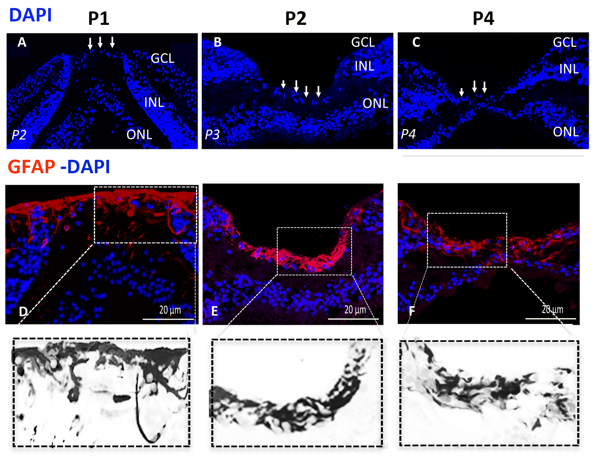
 Figure 1 of
Delaunay, Mol Vis 2020; 26:235-245.
Figure 1 of
Delaunay, Mol Vis 2020; 26:235-245.