Figure 3. Splicing results of minigene assays for three mutations in PAX6 in the HEK-293 or ARPE-19 cell lines. The schematics of the minigene constructs are depicted as follows: deep gray squares
= vectors, V = vector; light gray squares = PAX6 exons; thick lines = PAX6 introns; mutations are marked on the corresponding positions on the exons. PCR-primer2 and cDNA-primer1 were the primers
used for reverse transcription-polymerase chain reaction (RT-PCR) and sequencing. The bands labeled “*” represent an artifact
of the experimental process. A: RT–PCR analysis of minigene splicing for synonymous mutation c.174C>T and positive control c.357+1G>A in a transfected human
embryonic kidney 293 (HEK-293) cell line. Exon 5a was alternatively spliced in the wild-type and mutant constructs. The wild
type showed two splicing patterns: 629 bp (with exon5a)/587 bp and 428/386 bp, while mutation c.174C>T (M1) presented abnormal
splicing bands 444/402 bp that resulted in a 185 bp shortening of exon 6. The positive control c.357+1G>A (M2) also showed
abnormal splicing bands 521/479 bp that led to a 108 bp deletion of exon 6. (B) and (C). RT–PCR analysis of minigene splicing of missense mutations c.359T>A and c.780T>G in transfected HEK-293 and human retinal
pigmented epithelium-19 (ARPE-19) cell lines. Wild-type and mutant minigenes showed a normal splicing pattern, and no abnormal
splicing bands were observed. WT: wild-type; mut: mutant.
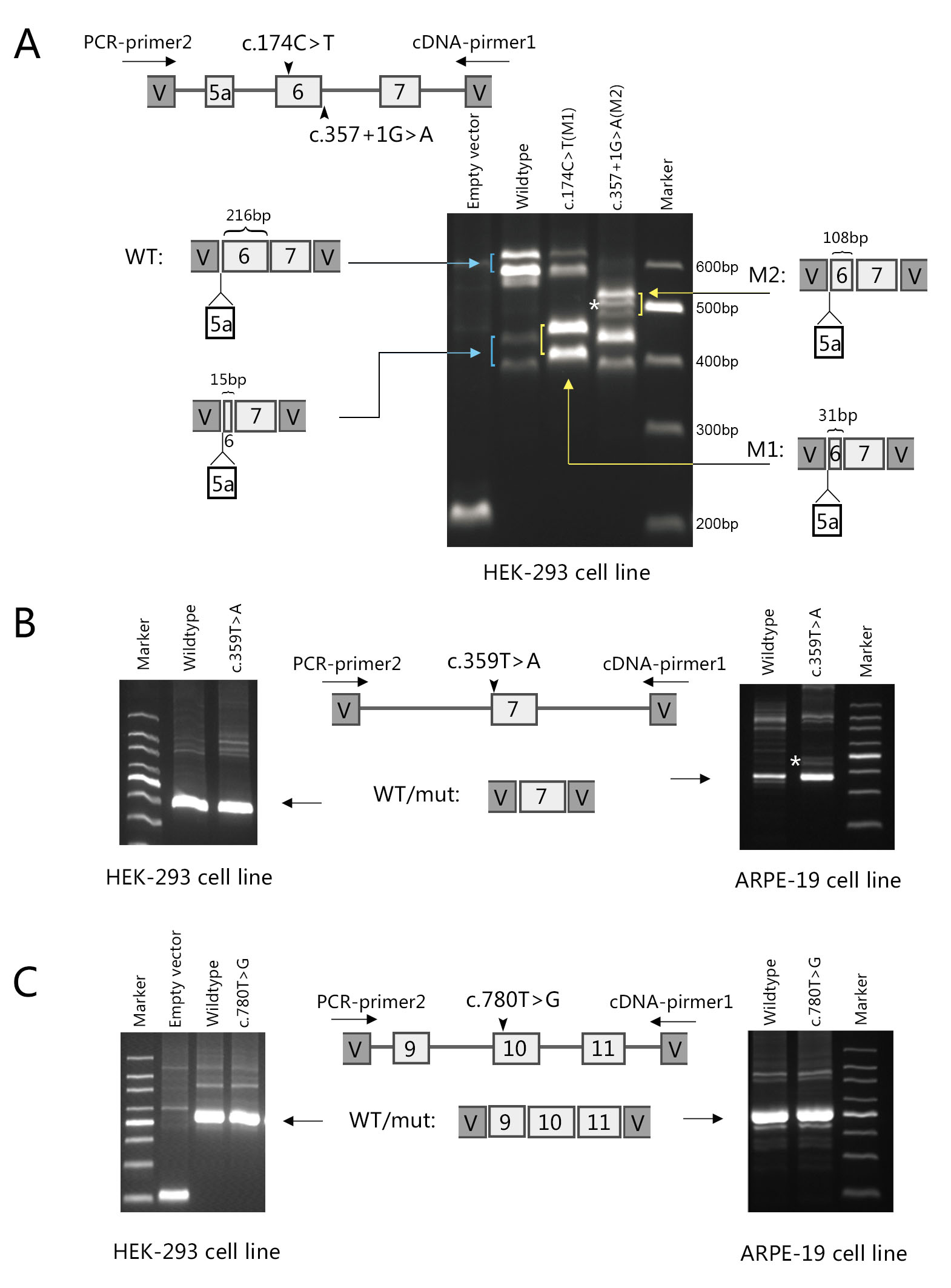
 Figure 3 of
You, Mol Vis 2020; 26:226-234.
Figure 3 of
You, Mol Vis 2020; 26:226-234.