Figure 2. The distribution of 58 distinct mutations in PAX6 detected in the present study. A: The distribution of 47 intragenic mutations on exons and introns of PAX6 and the corresponding domain of PAX6. LNK, HD, PD, and PST indicate the linker region, homeodomain, paired domain, and proline-threonine-serine-rich
domain, respectively. NMD, nonsense-mediated decay; IVS, intron. B: Lengths and positions of the 11 gross deletions involving PAX6 on chromosome 11p13. Cen indicates the centromere, and tel indicates the telomere. Gray and black squares indicate different
genes. Genes are shaded in different colors when they are near each other. The arrows indicate the coding direction of each
gene, and 026xxx indicates the patient ID.
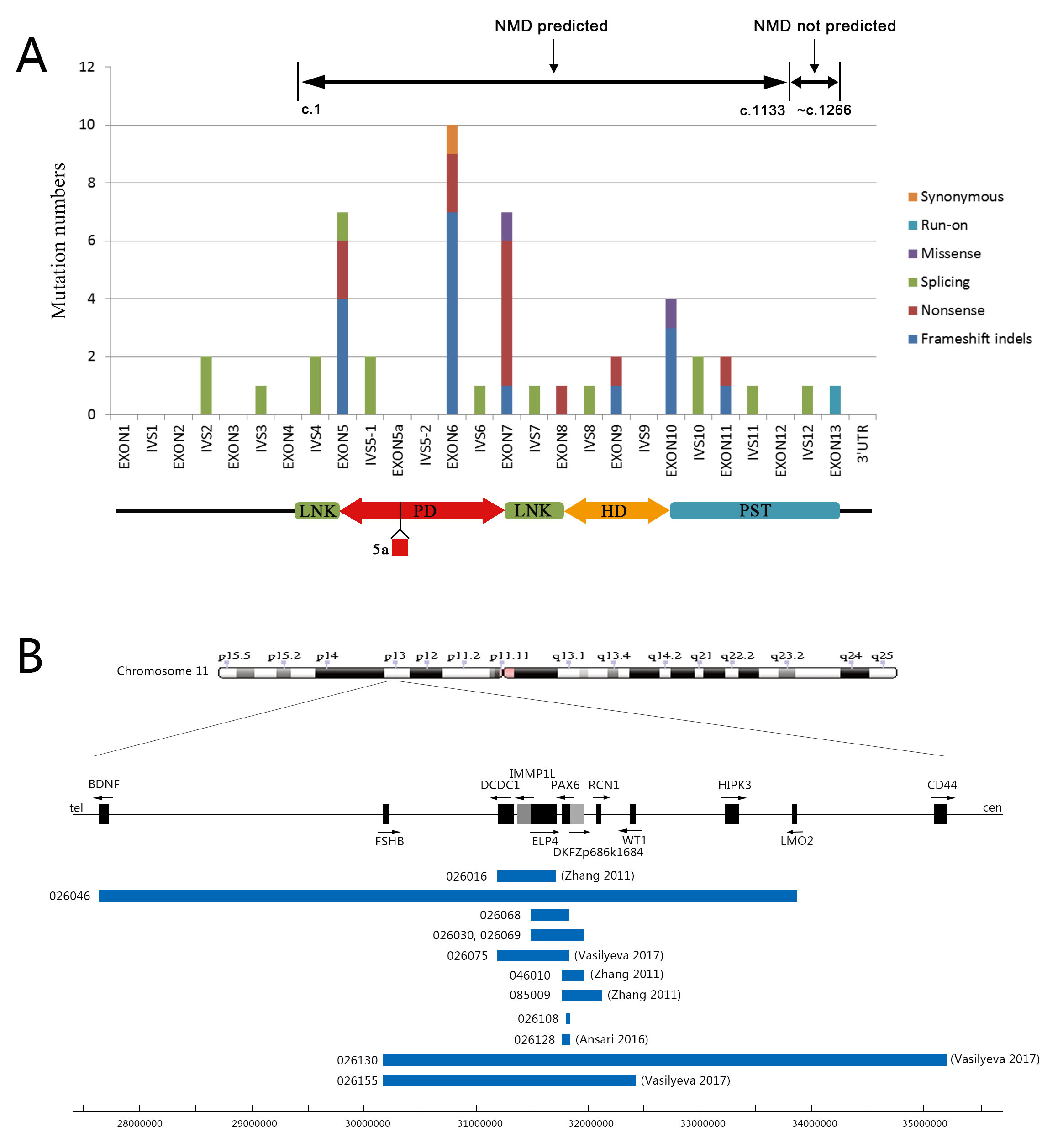
 Figure 2 of
You, Mol Vis 2020; 26:226-234.
Figure 2 of
You, Mol Vis 2020; 26:226-234.