Figure 4. Pedigrees of the families and segregation analysis of the detected variants. Arrows indicate the nucleotide position where
the variants are localized. Asterisks indicate individuals analyzed by whole exome sequencing in Family 1 and by clinical
exome in Family 2. wt: wild-type sequence.
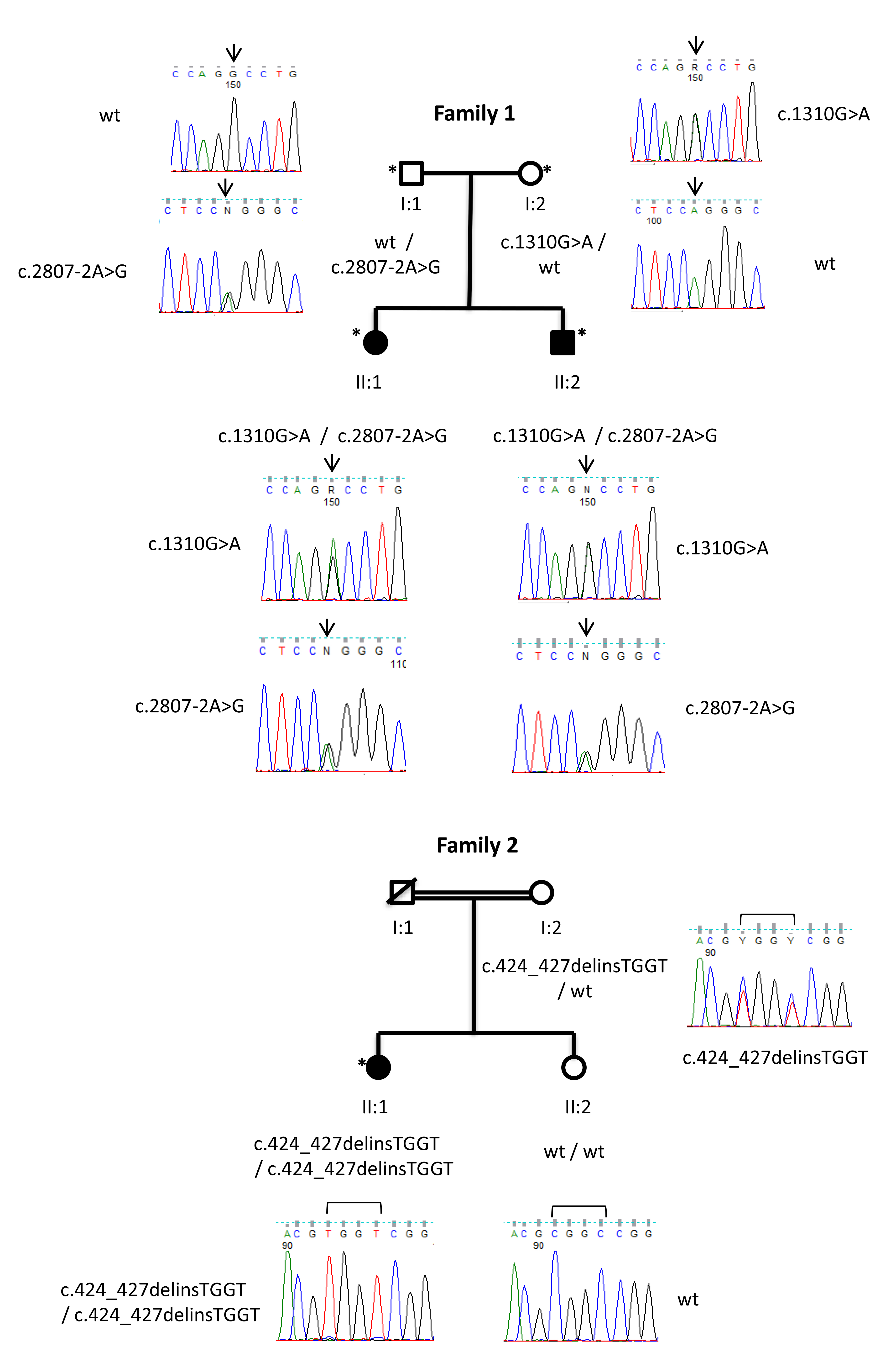
 Figure 4 of
García-García, Mol Vis 2020; 26:216-225.
Figure 4 of
García-García, Mol Vis 2020; 26:216-225.