Figure 5. Acute damage of retinal ganglion cells enhances β-catenin expression in Müller cells and gliosis reaction via Wnt/β-catenin
signaling in Müller cells. A, B: Western blot analyses (A) and densitometry (B) for β-catenin from retinal proteins of 8-week-old Slc1a3-CreERT/Ctnnb1fl/fl mice and Ctnnb1fl/fl controls 24 h after injection of N-methyl-D-aspartate (NMDA) or PBS into the vitreous cavity and 2 weeks after treatment
with tamoxifen (mean ± standard error of the mean, SEM; *p<0.05). C: Quantitative real-time RT–PCR for Gfap in RNA from retinas of 8-week-old Slc1a3-CreERT/Ctnnb1fl/fl mice and Ctnnb1fl/fl controls 7 h after injection of 3 µl of NMDA (10 mM) or PBS into the vitreous cavity and tamoxifen treatment 2 weeks before
(mean ± SEM; *p<0.05).
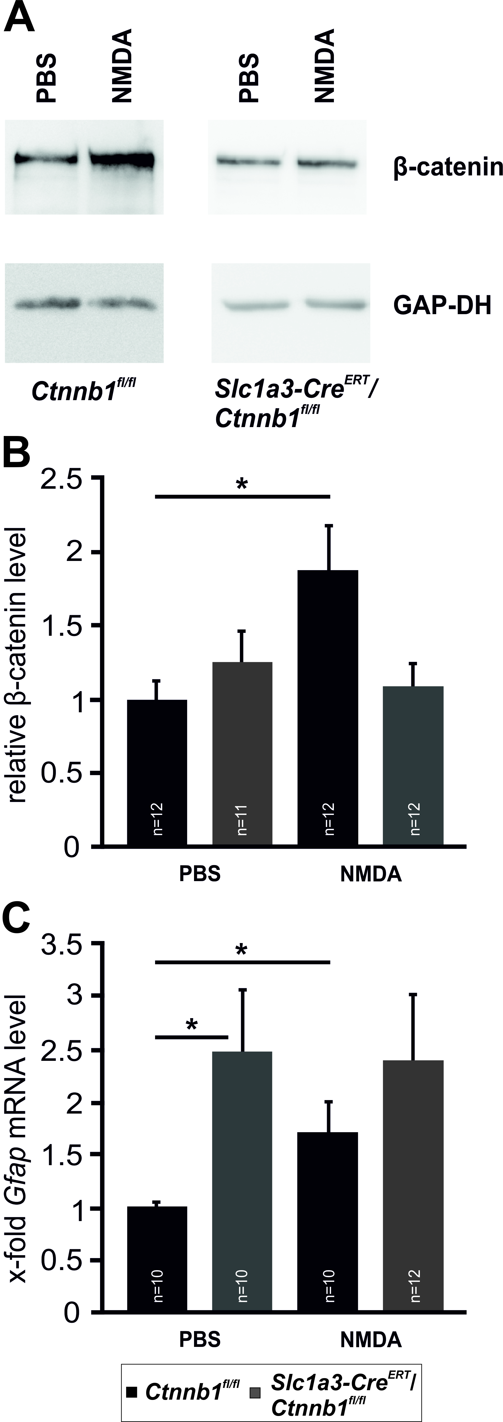
 Figure 5 of
Boesl, Mol Vis 2020; 26:135-149.
Figure 5 of
Boesl, Mol Vis 2020; 26:135-149.