Figure 4. Wnt/β-catenin deficiency in Müller cells amplifies apoptosis of retinal neurons. A: Representative terminal deoxynucleotidyl transferase dUTP nick-end labeling (TUNEL) staining of 8-week-old, tamoxifen-treated
Slc1a3-CreERT/Ctnnb1fl/fl mice and Ctnnb1fl/fl controls 24 h after injection of 3 µl of N-methyl-D-aspartate (NMDA; 10 mM) in one eye and PBS in the contralateral eye.
In the retinas of the Slc1a3-CreERT/Ctnnb1fl/fl mice, statistically significantly more TUNEL-positive cells in the retinal ganglion cell (RGC) layer were observed compared
to those in the Ctnnb1fl/fl controls. Intriguingly, no difference in the number of apoptotic cells was detected in the inner nuclear layer (INL) between
the Slc1a3-CreERT/Ctnnb1fl/fl and Ctnnb1fl/fl animals. Magnification bars, 50 µm; ONL, outer nuclear layer. B, C: The number of TUNEL-positive cells in the RGC layer (B) and the INL (C) was quantified and correlated to the retinal length (mean ± standard error of the mean, SEM; *p<0.05; ***p<0.001).
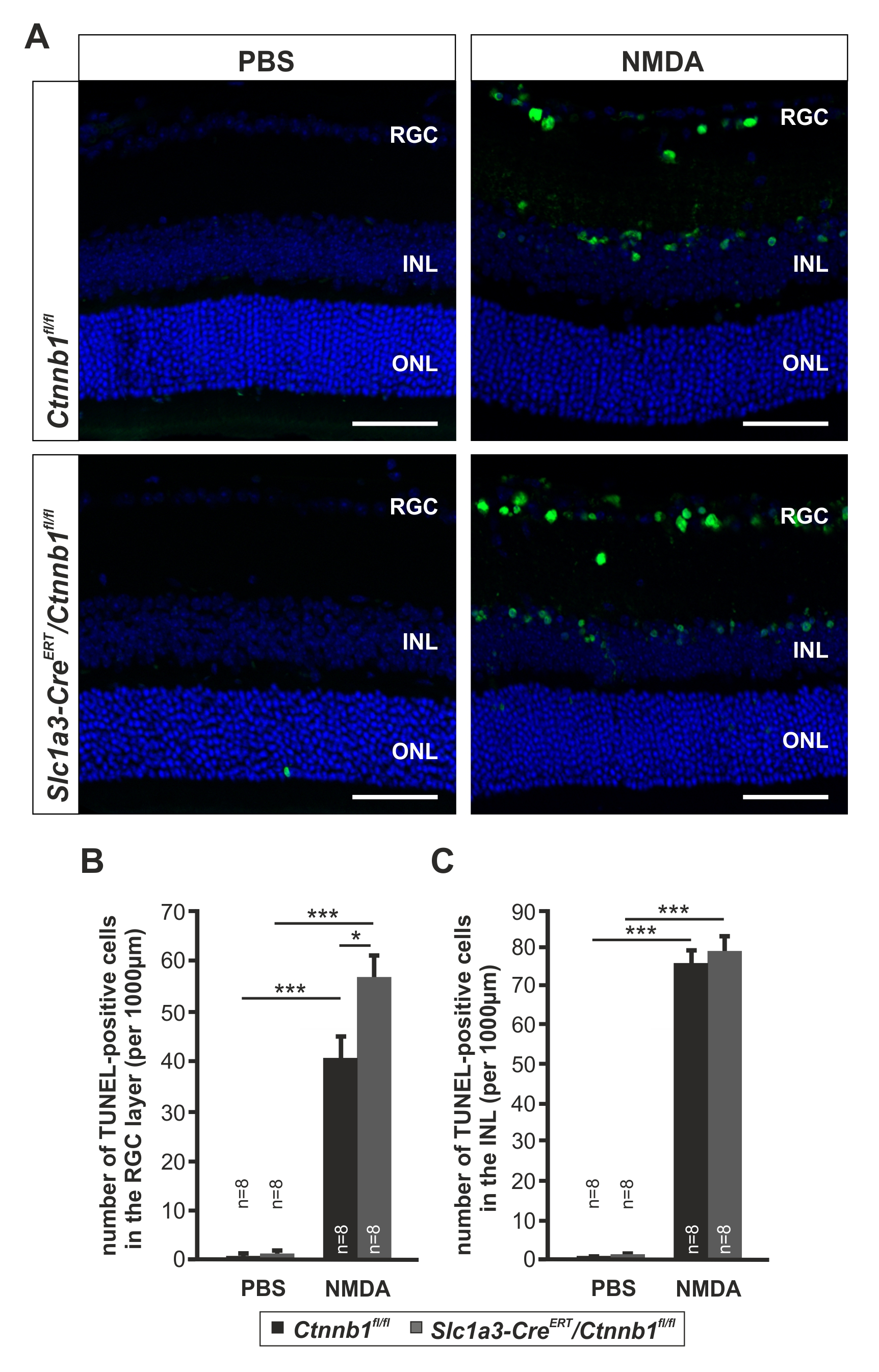
 Figure 4 of
Boesl, Mol Vis 2020; 26:135-149.
Figure 4 of
Boesl, Mol Vis 2020; 26:135-149.