Figure 3. Deficiency of Wnt/β-catenin signaling in Müller cells enhances degeneration of the inner retina following acute RGC damage.
A: Representative sagittal sections of retinas from 10-week-old Ctnnb1fl/fl and Slc1a3-CreERT/Ctnnb1fl/fl mice 2 weeks after injection of N-methyl-D-aspartate (NMDA) or PBS into the vitreous cavity and treatment with tamoxifen
at the age of 6 weeks. Scale bars, 50 µm. B–F: For quantification, the thickness of the (B) total retina, (C) the inner plexiform layer (IPL), (D) the inner nuclear layer (INL), (E) the outer plexiform layer (OPL), and (F) the outer nuclear layer (ONL) of 10-week-old Ctnnb1fl/f and Slc1a3-CreERT/Ctnnb1fl/fl mice 2 weeks after injection of PBS or NMDA into the vitreous cavity and treatment with tamoxifen 4 weeks before was measured
between every retinal tenth and plotted as a spider diagram. OS, ora serrata; ONH, optic nerve head; mean ± standard error
of the mean, SEM; n≥8; *p<0.05, **p<0.01, ***p<0.001; * indicates statistical significance between the two genotypes with
the same treatment; # (Ctnnb1fl/f) and + (Slc1a3-CreERT/Ctnnb1fl/fl) indicate statistical significance between NMDA and PBS-injected eyes.
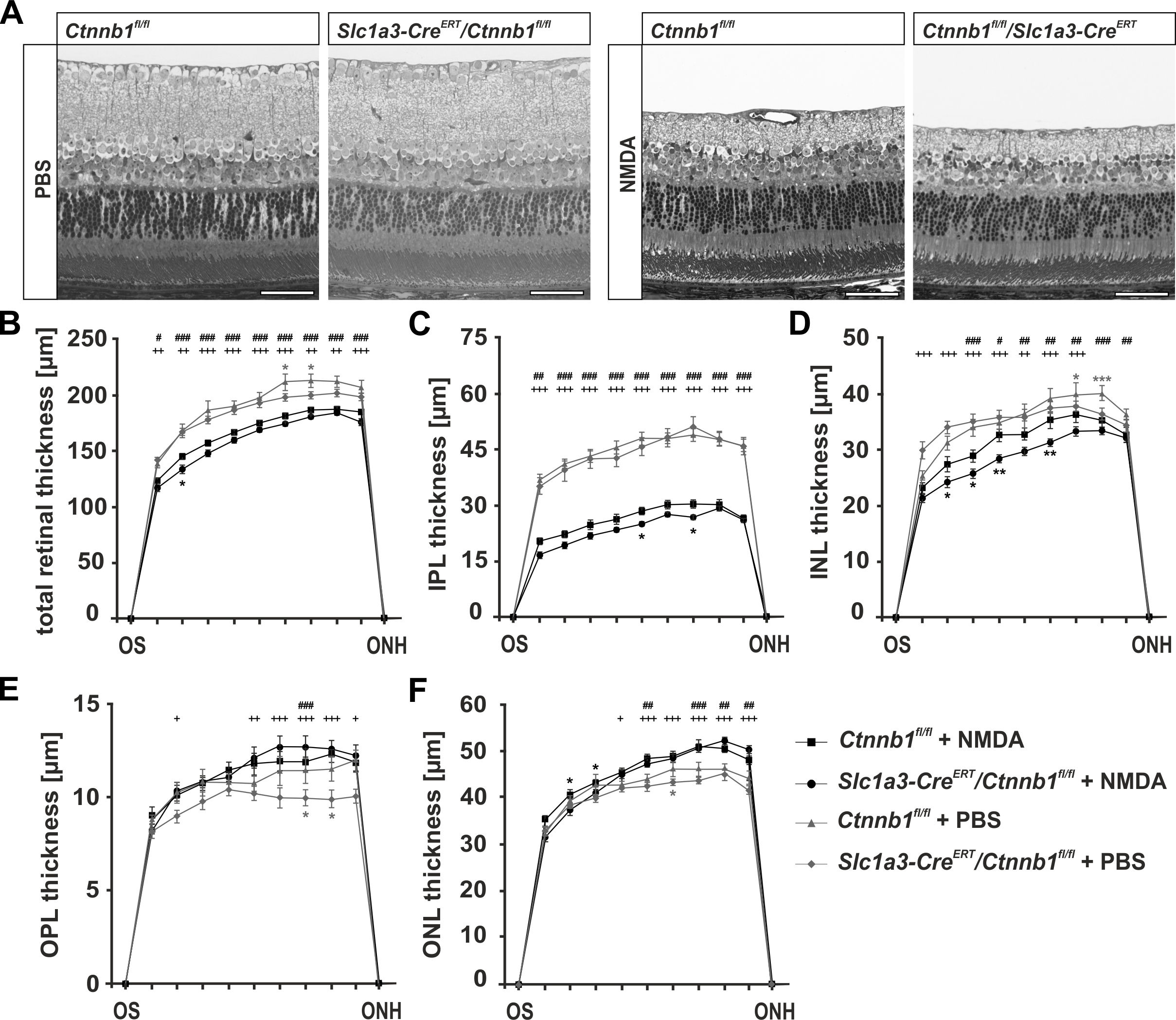
 Figure 3 of
Boesl, Mol Vis 2020; 26:135-149.
Figure 3 of
Boesl, Mol Vis 2020; 26:135-149.