Figure 2. Wnt/β-catenin deficiency in Müller cells enhances axon loss in the optic nerve following acute excitotoxic damage of RGCs.
A–H: Representative sagittal semithin sections through optic nerves from 10-week-old Slc1a3-CreERT/Ctnnb1fl/fl mice and Ctnnb1fl/fl controls 2 weeks after intravitreal injection of 3 µl N-methyl-D-aspartate (NMDA) [10 mM] or PBS and treatment with tamoxifen
at the age of 6 weeks (50 µl tamoxifen [20 mg/ml] i.p. 2x/day for 5 days). In the optic nerves of the NMDA-injected eyes,
obvious loss of axons, broad glial scars (asterisk), and darkly stained myelin whirls (arrow heads) were observed (E–H), whereas in the PBS-treated eyes only a few degenerating myelin sheathes were detectable (A–D). In the Slc1a3-CreERT/Ctnnb1fl/fl mice, the axonal damage was even more pronounced compared to that in the Ctnnb1fl/fl controls (E–H). Magnification bars: A, C, E, G, 50 µm; B, D, F, H, 10 µm. I: For quantification, the number of axons in the optic nerves from the Slc1a3-CreERT/Ctnnb1fl/fl mice and the Ctnnb1fl/fl controls was quantified and plotted as the total number per optic nerve (mean ± standard error of the mean, SEM; *p<0.05;
***p<0.001).
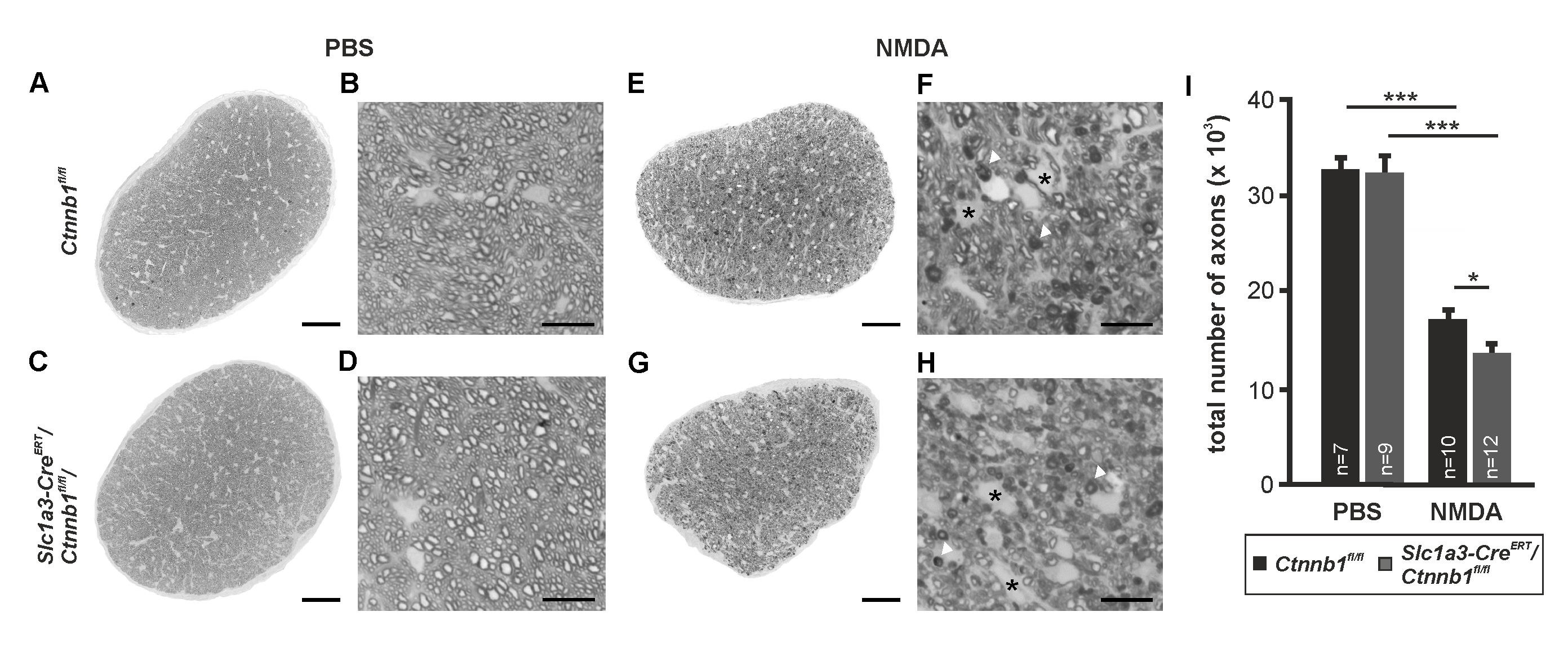
 Figure 2 of
Boesl, Mol Vis 2020; 26:135-149.
Figure 2 of
Boesl, Mol Vis 2020; 26:135-149.