Figure 1. Tamoxifen-induced β-catenin deficiency in Müller cells of Slc1a3-CreERT/Ctnnb1fl/fl mice. A, B: Representative β-galactosidase staining of 8-week-old Rosa26-LacZ reporter mice with or without additional Cre expression
in Müller cells (Slc1a3-CreERT) after treatment with tamoxifen (50 µl tamoxifen [20 mg/ml] i.p. 2x/day for 5 days). In the Slc1a3-CreERT/Rosa26-LacZ mice, radial staining for β-galactosidase that spread from the inner to the outer limiting membrane was detected.
In addition, the nuclei of the labeled cells were localized in the inner nuclear layer (B), whereas no β-galactosidase expression was detected in mice without Cre expression (A). Magnification bars = 50 µm. RGC, retinal ganglion cell, INL, inner nuclear layer; ONL, outer nuclear layer. C, D: Western blot analysis (C) and densitometry (D) for β-catenin from retinal proteins of 8-week-old Slc1a3-CreERT/Ctnnb1fl/fl mice 2 weeks after treatment with tamoxifen (mean ± standard error of the mean, SEM; n = 8; *p<0.05). E–G: Real-time RT–PCR (E) and immunohistochemical staining (F, G) for Sox9 of 8-week-old mice 2 weeks after treatment with tamoxifen (50 µl tamoxifen [20 mg/ml] i.p. 2x/day for 5 days).
No difference in the number or distribution of Sox9 positive nuclei (red) in the INL was seen between the Ctnnb1fl/fl (F) and Slc1a3-CreERT/Ctnnb1fl/fl (G) mice. Magnification bars, 50 µm. Blue, 4′,6-diamidino-2-phenylindole (DAPI) staining; RGC, retinal ganglion cell, INL, inner
nuclear layer; ONL, outer nuclear layer.
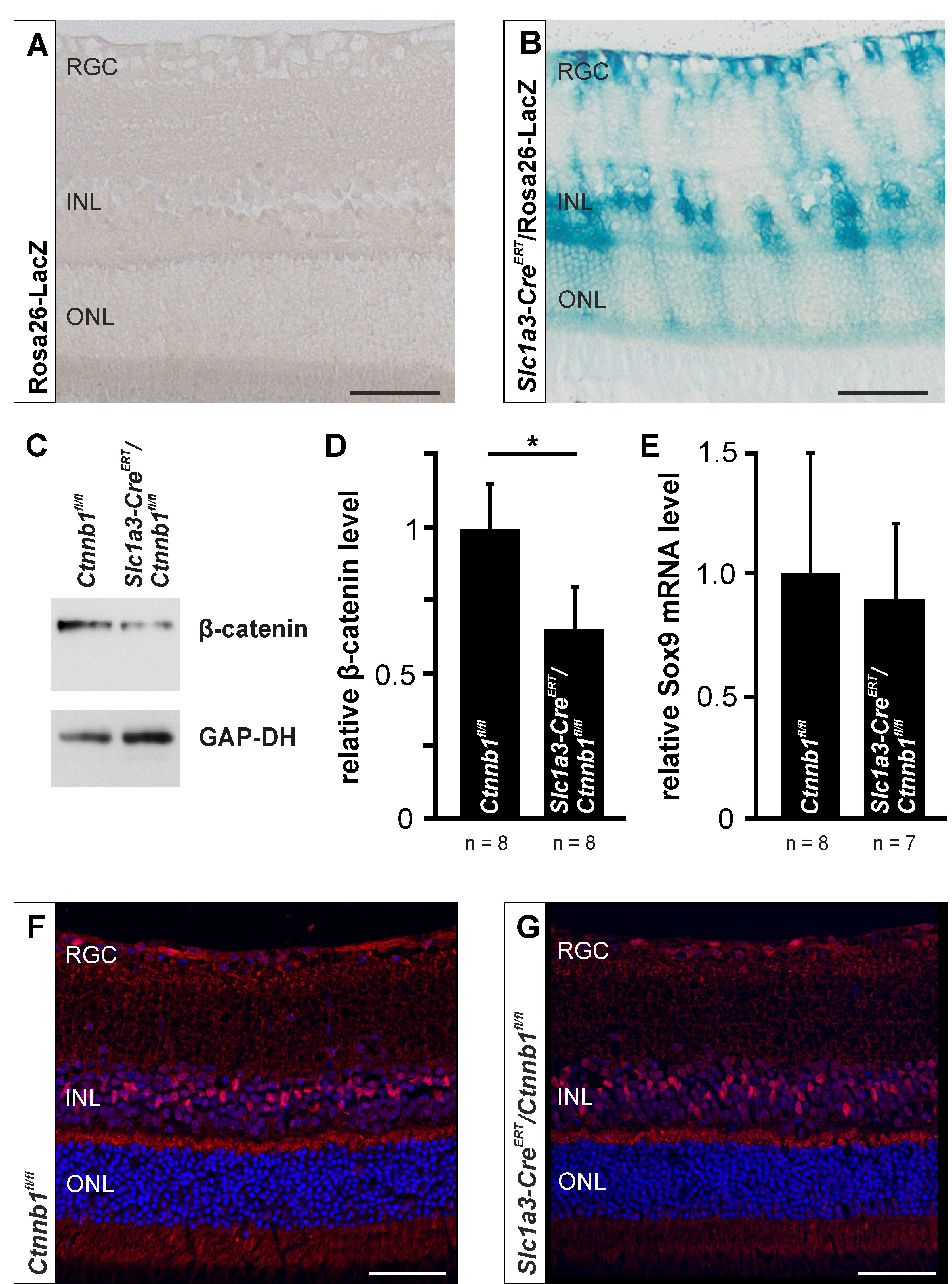
 Figure 1 of
Boesl, Mol Vis 2020; 26:135-149.
Figure 1 of
Boesl, Mol Vis 2020; 26:135-149.