Figure 6. Biochemical analysis of GCAP1-D144G. A: Limited proteolysis of GCAP1-wild type (WT) and GCAP1-D144G by trypsin in the presence and absence of Ca2+. Digestions were performed at 30 °C for 0, 5, 10, and 20 min. B: Electrophoresis shift assay of GCAP1-WT and GCAP1-D144G in the presence of 2.5 μM Ca2+. C: The expression level and stability of GCAP1-WT and GCAP1-D144G in human embryonic kidney (HEK)-293 cells. GCAP1-WT and GCAP1-D144G
were transfected into the HEK-293 cells. The proteins were detected using anti-FLAG antibody at 24, 48, and 72 h. The bars
and error bars represent the mean ± standard deviation (SD) of three independent experiments, and the values within each experiment
were normalized to those of glyceraldehyde-3-phosphate dehydrogenase (GAPDH). CONT, control.
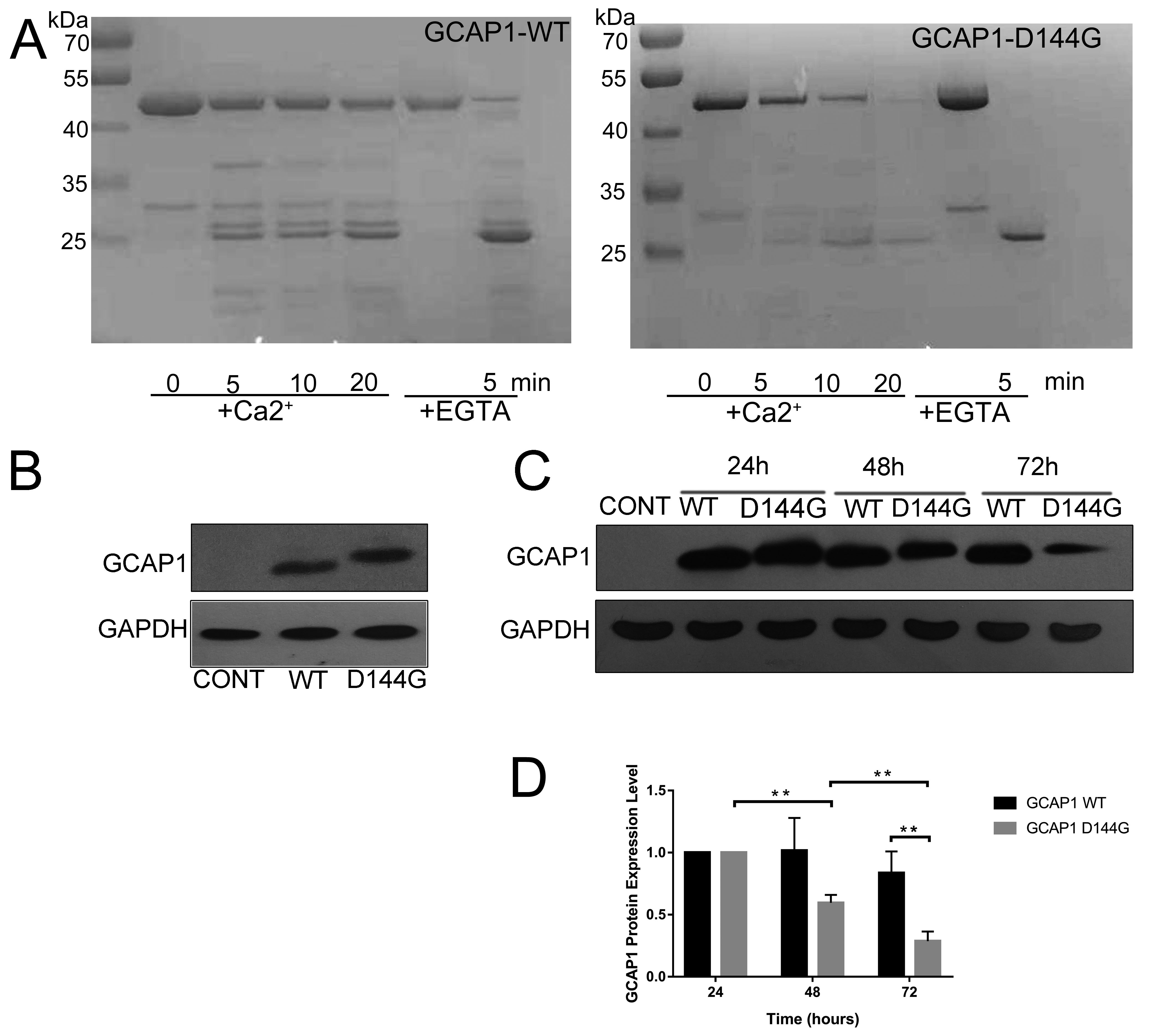
 Figure 6 of
Tang, Mol Vis 2019; 25:921-933.
Figure 6 of
Tang, Mol Vis 2019; 25:921-933.