Figure 3. Phenotypic appearance of patient IV:3. A: Both eyes have a clear optic disc boundary and a slightly dark macular area. The left eye shows diffuse depigmentation (window-like
defect) and generally normal retinal vessels, with no bleeding or exudation in the retina. B: A slightly high reflex zone is visible on the temporal lateral of the central macular fovea in the left eye on infrared
fundus photography. C: The macular optical coherence tomography (OCT) examination shows that the band imaging of both eyes is slightly blurred.
D: Multifocal electroretinography indicates that the amplitude density of the macular area in the left eye is significantly
reduced. E: The Humphrey visual field examination disclosed that the visual field of both eyes has defects in different degrees.
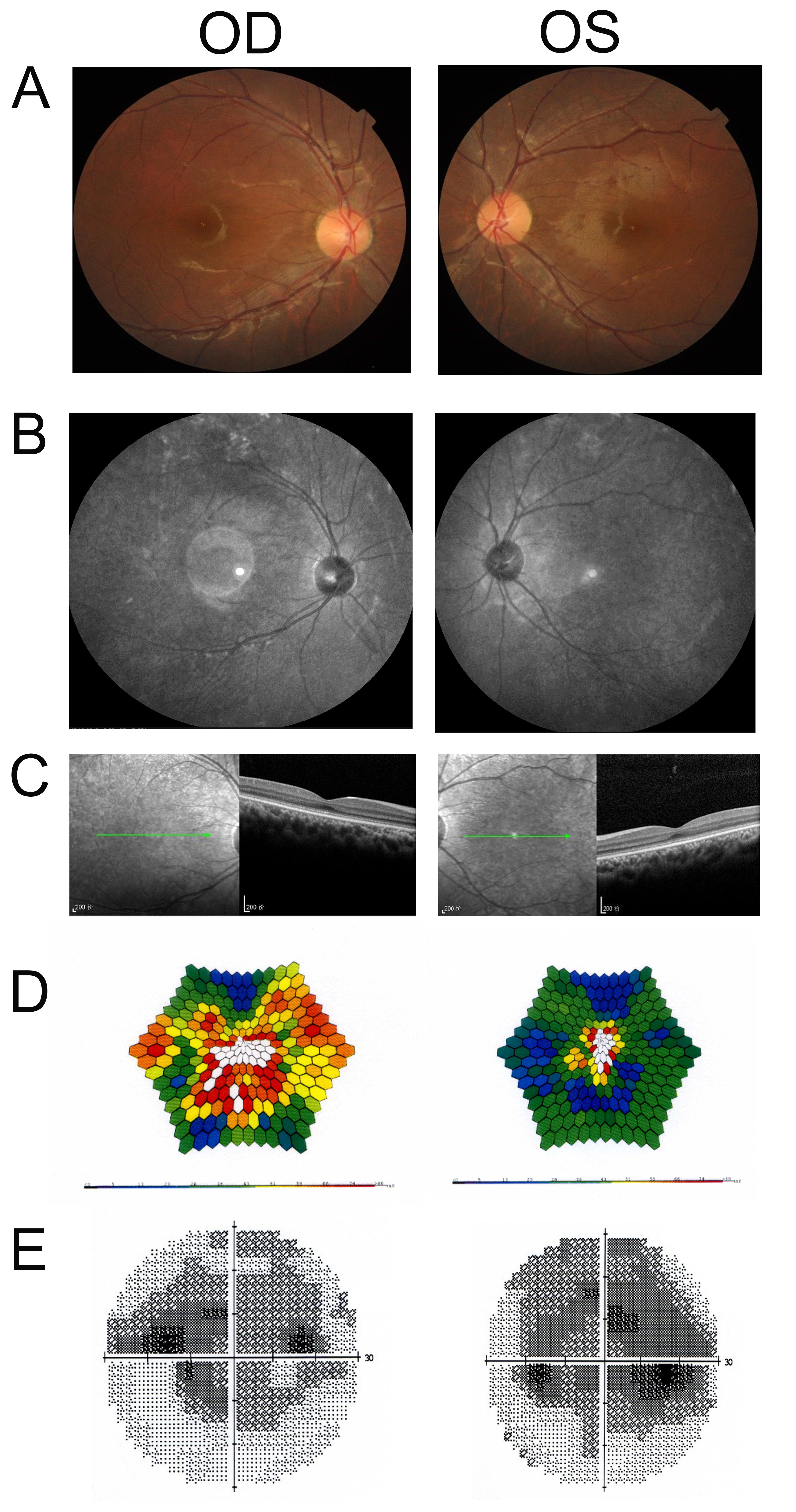
 Figure 3 of
Tang, Mol Vis 2019; 25:921-933.
Figure 3 of
Tang, Mol Vis 2019; 25:921-933.