Figure 2. Phenotypic appearance of the COD proband (III:2). A: The fundus photograph shows that III:2 had oval lesions in the macular area and discoid depigmentation. The foveal reflex
disappeared in both eyes. The macular area of her left eye is blue-gray with golden foil reflections, and the boundary is
clear. There is irregular, patchy hyperplasia in her left eye. B: The infrared fundus photograph demonstrates the depigmentation zone in the macular area and slightly higher reflection in
the mid-peripheral area. C: On the macular optical coherence tomography (OCT) images, the foveal centralis of both eyes is thin, and the ellipsoid and
chimera bands are absent. Subepithelial hyper-reflexes are deposited, and choroidal capillary layer reflexes are enhanced
in the foveal centralis. OD (right eye), OS (left eye).
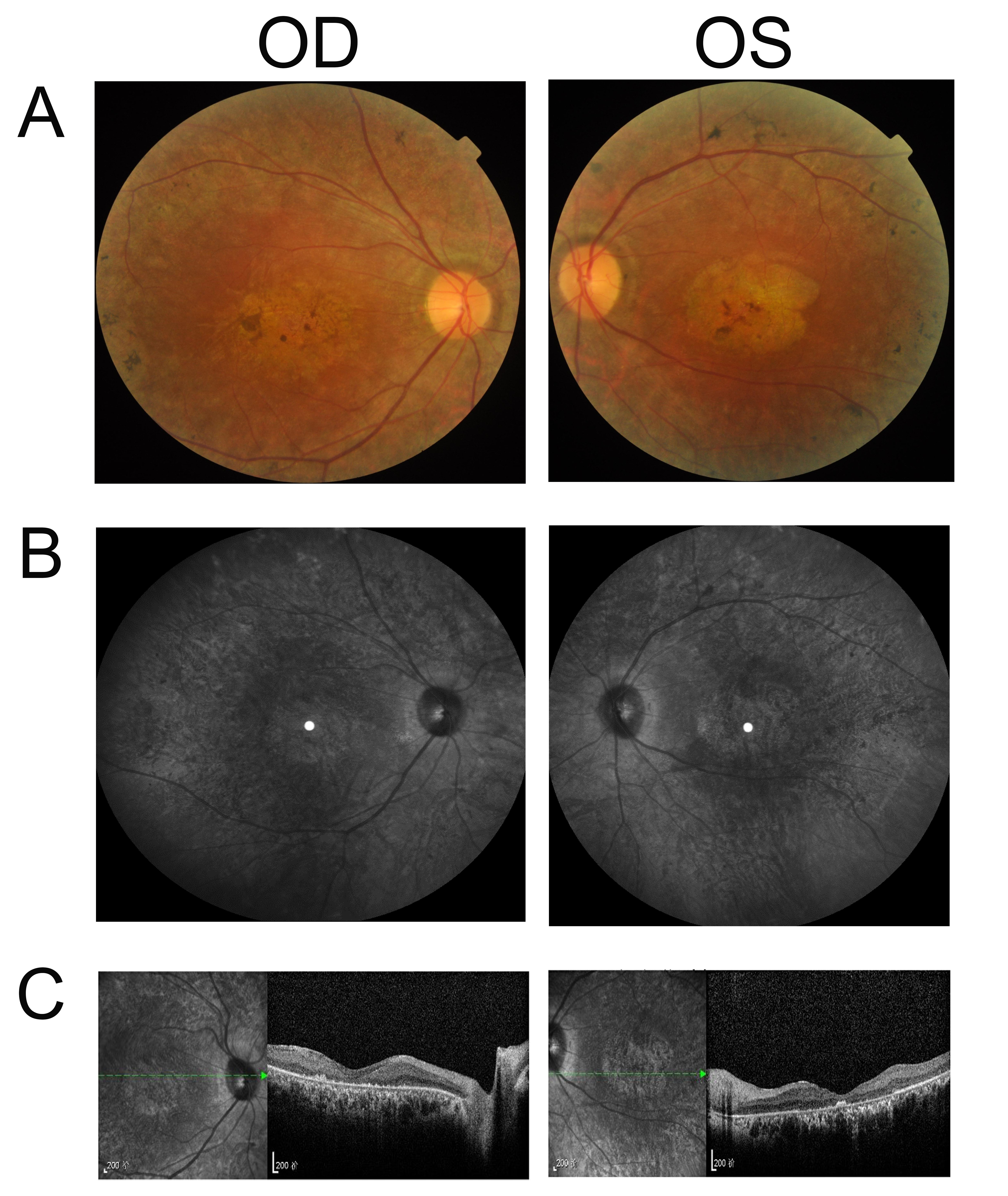
 Figure 2 of
Tang, Mol Vis 2019; 25:921-933.
Figure 2 of
Tang, Mol Vis 2019; 25:921-933.