Figure 2 of
Wang, Mol Vis 2019; 25:912-920.
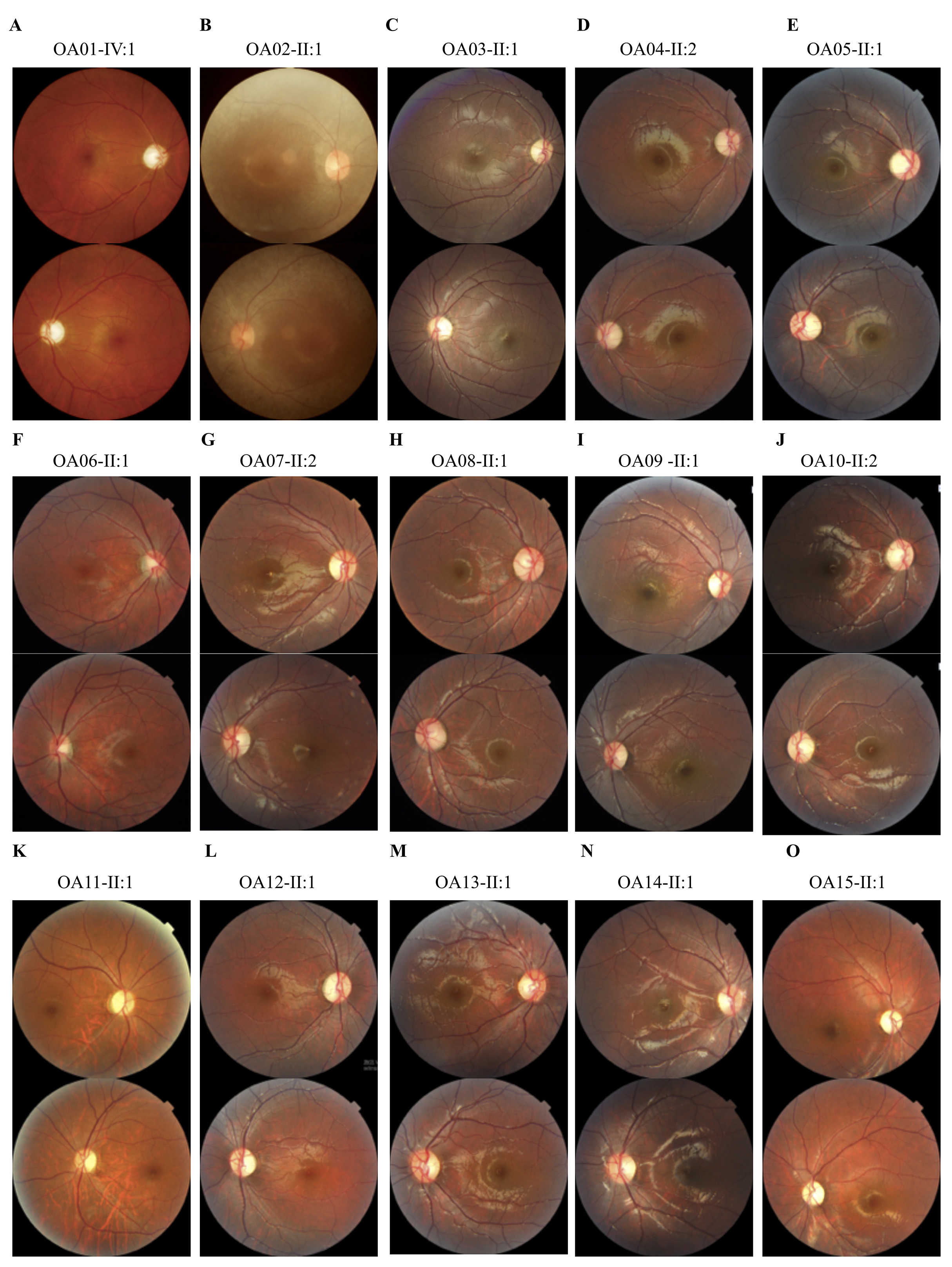
Figure 2.
Fundus presentations.
A-O
: Fundus photographs of all probands demonstrate bilateral optic nerve head pallor.
 Figure 2 of
Wang, Mol Vis 2019; 25:912-920. Figure 2 of
Wang, Mol Vis 2019; 25:912-920.
Figure 2 of
Wang, Mol Vis 2019; 25:912-920. Figure 2 of
Wang, Mol Vis 2019; 25:912-920.