Figure 6. Longitudinal evaluation of SD-OCT image in adult Grm6nob8 mice. A: The upper image is a representative image taken across the horizontal meridian from a Grm6nob8 mouse at 57 weeks of age showing the well-defined laminar structure of the retina. Lower images were taken from Grm6nob8 mice at the ages indicated. ELM, external limiting membrane; EZ, ellipsoid zone; INL, inner nuclear layer; IPL, inner plexiform
layer; ONL, outer nuclear layer; OPL, outer plexiform layer; RPE, retinal pigment epithelium. B: Thickness of the INL at 11, 39, and 57 weeks of age. The INL was thicker at 11 weeks of age, compared to the older ages
(Aspin-Welch’s t test; *p<0.05, **p<0.01). Each plot indicates average +/− standard error of four (11 weeks old) or three (39 and 57 weeks
old) mice.
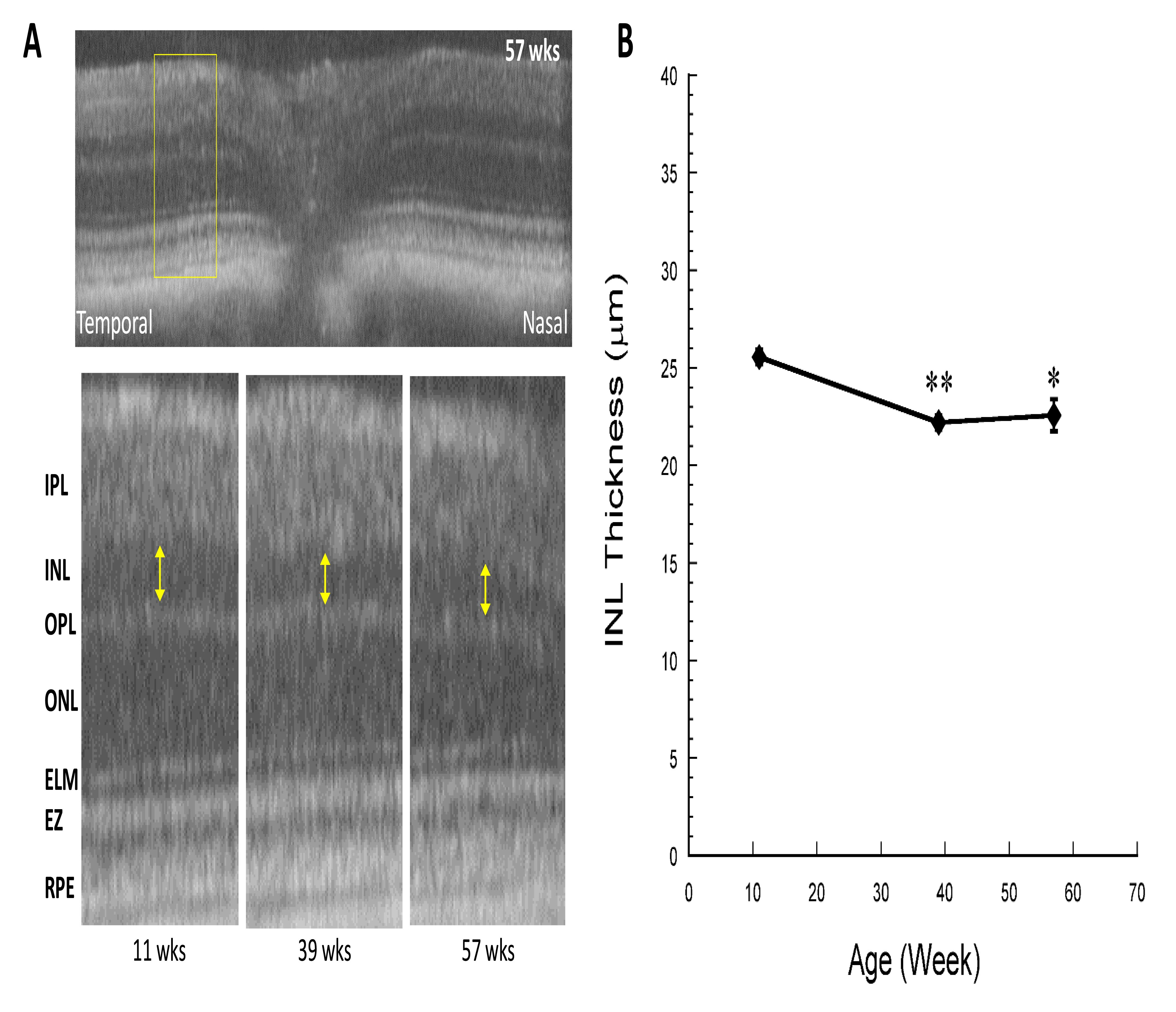
 Figure 6 of
Kinoshita, Mol Vis 2019; 25:890-901.
Figure 6 of
Kinoshita, Mol Vis 2019; 25:890-901.