Figure 1. Western blotting analysis of mGluR6 content in WT and Grm6 mutants. A: Western blots for mGluR6 in wild-type (WT; lanes 1 and 2), Grm6nob8 homozygotes (lanes 3 and 4), Grm6nob3/nob8 compound heterozygotes (lanes 5 and 7), and a Grm6nob3 homozygote (lane 6). B: Densitometric analysis of western blots, demonstrating that mGluR6 levels are dramatically reduced in all Grm6 mutants, and that mGlur6 levels of Grm6nob3/nob8 compound heterozygotes are reduced to approximately half of the levels seen in Grm6nob8 homozygotes. Each bar indicates average +/− standard error of three samples. Statistically significant differences were detected
between groups with Aspin-Welch’s t test (*p<0.05, **p<0.01).
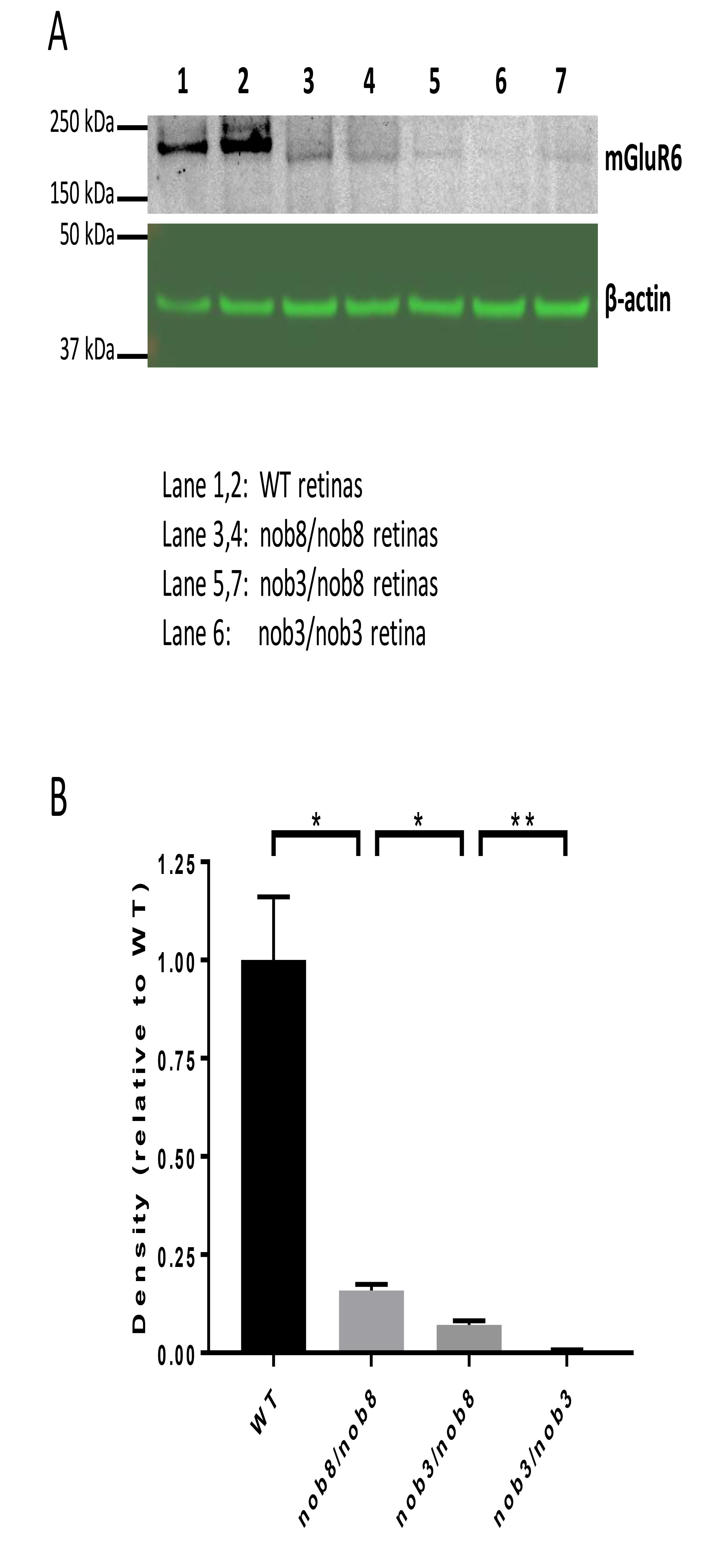
 Figure 1 of
Kinoshita, Mol Vis 2019; 25:890-901.
Figure 1 of
Kinoshita, Mol Vis 2019; 25:890-901.