Figure 9. Clinical imagining of patient 9 with a c.68C>A mutation in RHO (p.Pro23His). A, C: Oculus dexter (OD) color photos showing scant bone spicules in the inferotemporal periphery. B, D: Oculus sinister (OS) color photos showing scant bone spicules in inferotemporal periphery. E: OD fundus autofluorescence (FAF) fundus photos showing a crescent-shaped area of hypoautofluorescence (hypoAF) along the
inferior arcades extending into the inferior midperiphery as well as the infero- and superonasal periphery. F: OS FAF fundus photos showing a crescent-shaped area of hypoAF along the inferior arcades extending into the inferior midperiphery
as well as the infero- and superonasal periphery. G: OD foveal spectral-domain optical coherence tomography (SD-OCT) showing mild outer retinal attenuation. H: OS foveal SD-OCT showing mild outer retinal attenuation. I: OS Goldman visual field showing circumferential constriction. J: OD Goldman visual field showing circumferential constriction.
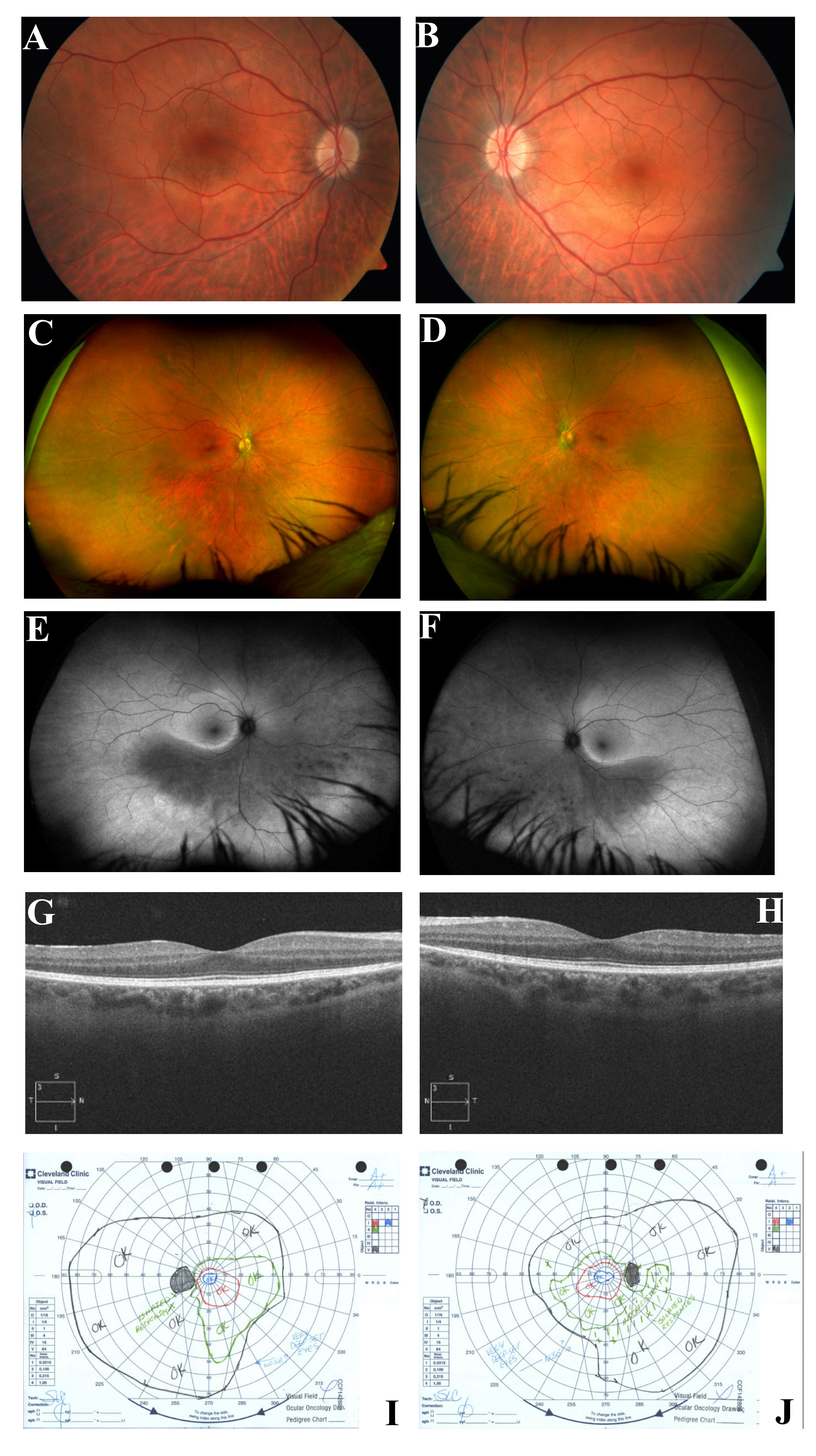
 Figure 9 of
Coussa, Mol Vis 2019; 25:869-889.
Figure 9 of
Coussa, Mol Vis 2019; 25:869-889.