Figure 8. Clinical imagining of patient 8 with a c.325G>A mutation in RHO (p.Gly109Arg). A, E: Oculus dexter (OD) color photos showing RPE hypopigmentation and atrophic changes extending inferiorly from the inferior
arcade border to the periphery. B, F: Oculus sinister (OS) color photos showing RP RPE hypopigmentation and atrophic changes extending inferiorly from the inferior
arcade border to the periphery. C, G: OD fundus autofluorescence (FAF) fundus photos showing a linear hyper-autofluorescence (hyperAF) abutting the inferior edge
of the fovea. An OU patchy pattern of hypoautofluorescence (hypoAF) extending from the inferior border of the inferior arcade
into the inferior and inferonasal midperiphery. D, H: OS FAF fundus photos showing linear hyperAF abutting the inferior edge of the fovea. OU patchy pattern of hypoAF extending
from the inferior border of the inferior arcade into the inferior and inferonasal midperiphery. I: OD spectral-domain optical coherence tomography (SD-OCT) of the superior posterior pole showing retinal thinning and ellipsoid
zone loss of the superior posterior pole adjacent to the arcades. J: OD foveal SD-OCT showing a small island of preservation in the ellipsoid zone subfoveally with parafoveal loss of the ellipsoid
zone. K: OD SD-OCT of the inferior posterior pole showing marked retinal disorganization and RPE hyper-reflective round deposits
adjacent to the arcades with significant choroidal hyporeflective signal. L: OS SD-OCT of the superior posterior pole showing retinal thinning and ellipsoid zone loss adjacent to the arcades. M: OS foveal SD-OCT showing a small island of preservation in the ellipsoid zone subfoveally with parafoveal loss of the ellipsoid
zone. N: OS SD-OCT of the inferior posterior pole showing marked retinal disorganization and RPE hyper-reflective round deposits
adjacent to the arcades with significant choroidal hyporeflective signal. O: OS Goldman visual field showing mild nasal constriction. P: OD Goldman visual field showing small superior defects and mild temporal constriction.
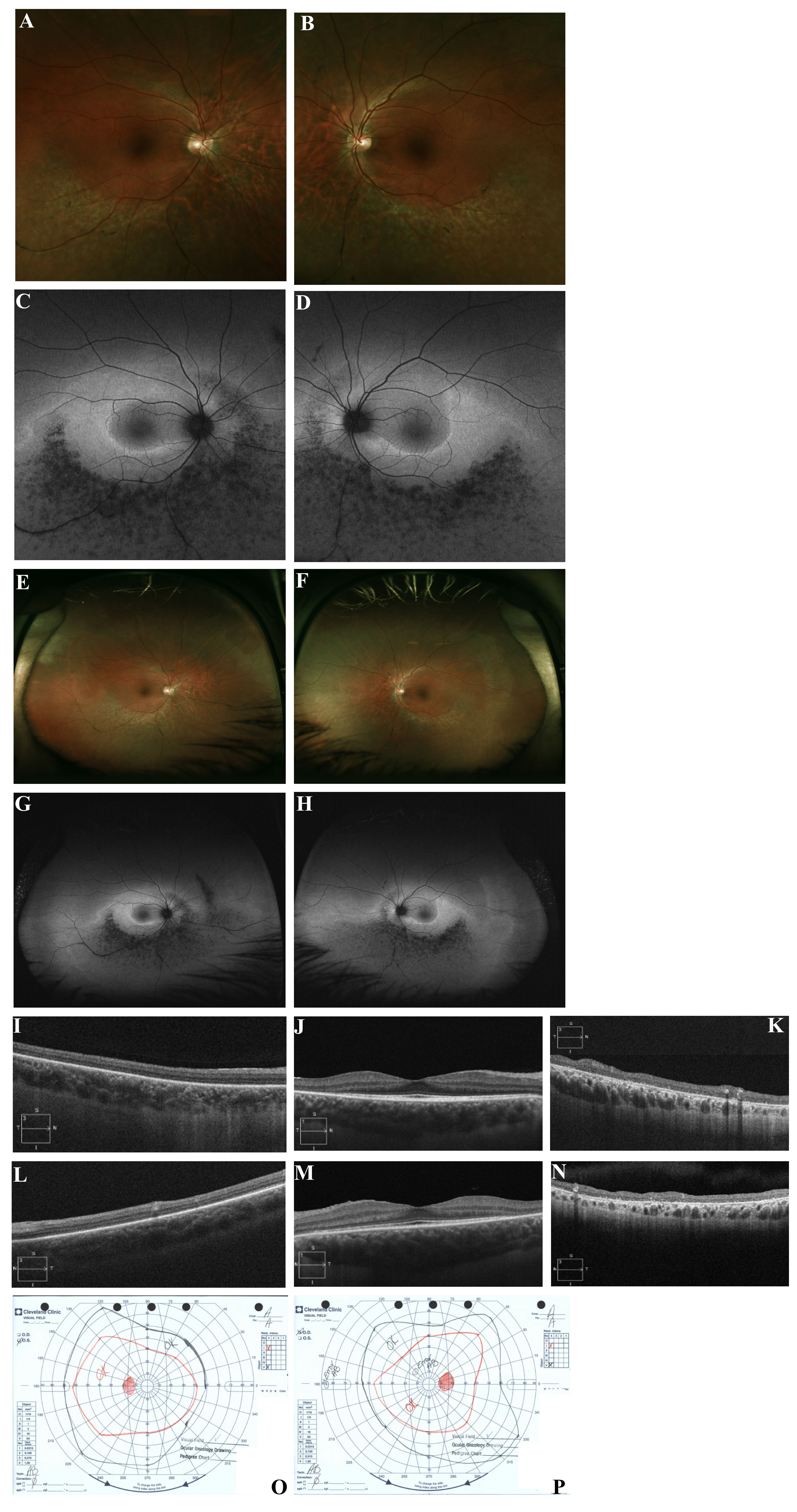
 Figure 8 of
Coussa, Mol Vis 2019; 25:869-889.
Figure 8 of
Coussa, Mol Vis 2019; 25:869-889.