Figure 7. Clinical imagining of patient 7 with a c.44A>G mutation in RHO (p.Asn15Ser). A, E: Oculus dexter (OD) color photos showing a concentric foveal hypopigmentation, marked parafoveal RPE mottling and hypopigmentation,
RPE atrophy, and bone spicules adjacent to and along the inferior arcades with mild extension temporally to the fovea as well
as temporally and superonasally with respect to the optic disc and superonasally with respect to the superior arcade. B, F: Oculus sinister (OS) color photos showing concentric foveal hypopigmentation, marked parafoveal RPE mottling and hypopigmentation,
RPE atrophy, and bone spicules adjacent to and along the inferior arcades with mild extension temporally to the fovea as well
as temporally and superonasally with respect to the optic disc and superonasally with respect to the superior arcade. C, G: OD fundus autofluorescence (FAF) fundus photos showing a central hyper-autofluorescence (hyperAF) rim surrounded by a speckled
pattern of hypoautofluorescence (hypoAF) forming a “bull’s eye”-like pattern. There is also a circinate area of hypoAF along
the inferior and superior arcades with a patchy pattern of marked hypoAF extending inferiorly from the inferior arcade into
the periphery. D, H: OS FAF fundus photos showing a central hyperAF rim surrounded by a speckled pattern of hypoAF forming a “bull’s eye”-like
pattern. There is also a circinate area of hypoAF along the inferior and superior arcades with a patchy pattern of marked
hypoAF extending inferiorly from the inferior arcade into the periphery. I: OD foveal spectral-domain optical coherence tomography (SD-OCT) showing a small island of preservation in the ellipsoid
zone subfoveally, marked retinal layers contour abnormalities and thinning, RPE hyperreflective round deposits, significant
choroidal hyper-reflective signal, and intraretinal cystic spaces. J: OS foveal SD-OCT showing a small island of preservation in the ellipsoid zone subfoveally, marked retinal layer contour
abnormalities and thinning, RPE hyperreflective round deposits, significant choroidal hyper-reflective signal, and intraretinal
cystic spaces. K: OS Goldman visual field showing superonasal and inferonasal defects. L: OD Goldman visual field showing superonasal defects.
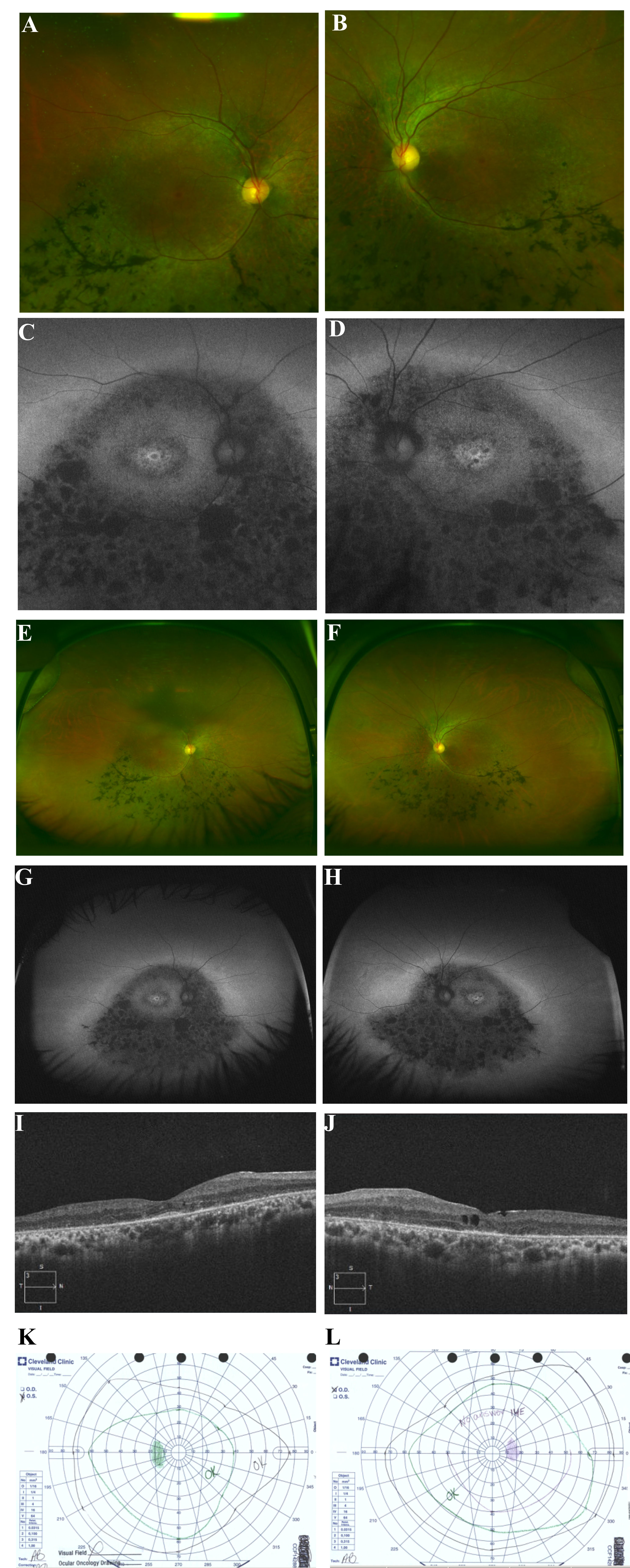
 Figure 7 of
Coussa, Mol Vis 2019; 25:869-889.
Figure 7 of
Coussa, Mol Vis 2019; 25:869-889.