Figure 6. Clinical imagining of patient 6 with a c.44A>G mutation in RHO (p.Asn15Ser). A, C: Oculus dexter (OD) color photos of the posterior pole showing marked RPE atrophy and bone spicules adjacent to and along
the inferior arcades with mild extension temporally to the fovea as well as superonasally with respect to the optic disc and
the superior arcade. B, D: Oculus sinister (OS) color photos of the posterior pole showing marked RPE atrophy and bone spicules adjacent to and along
the inferior arcades with mild extension temporally to the fovea as well as superonasally with respect to the optic disc and
the superior arcade. E: OD foveal spectral-domain optical coherence tomography (SD-OCT) showing parafoveal retinal thinning and loss of the ellipsoid
zone and photoreceptor layer most prominent in the inferior part of the posterior pole. F: OS foveal SD-OCT showing parafoveal retinal thinning and loss of the ellipsoid zone and photoreceptor layer most prominent
in the inferior part of the posterior pole. G: OS Humphrey 30-2 SITA Standard visual field showing circumferential constriction. H: OD Humphrey 30-2 SITA Standard visual field showing circumferential constriction.
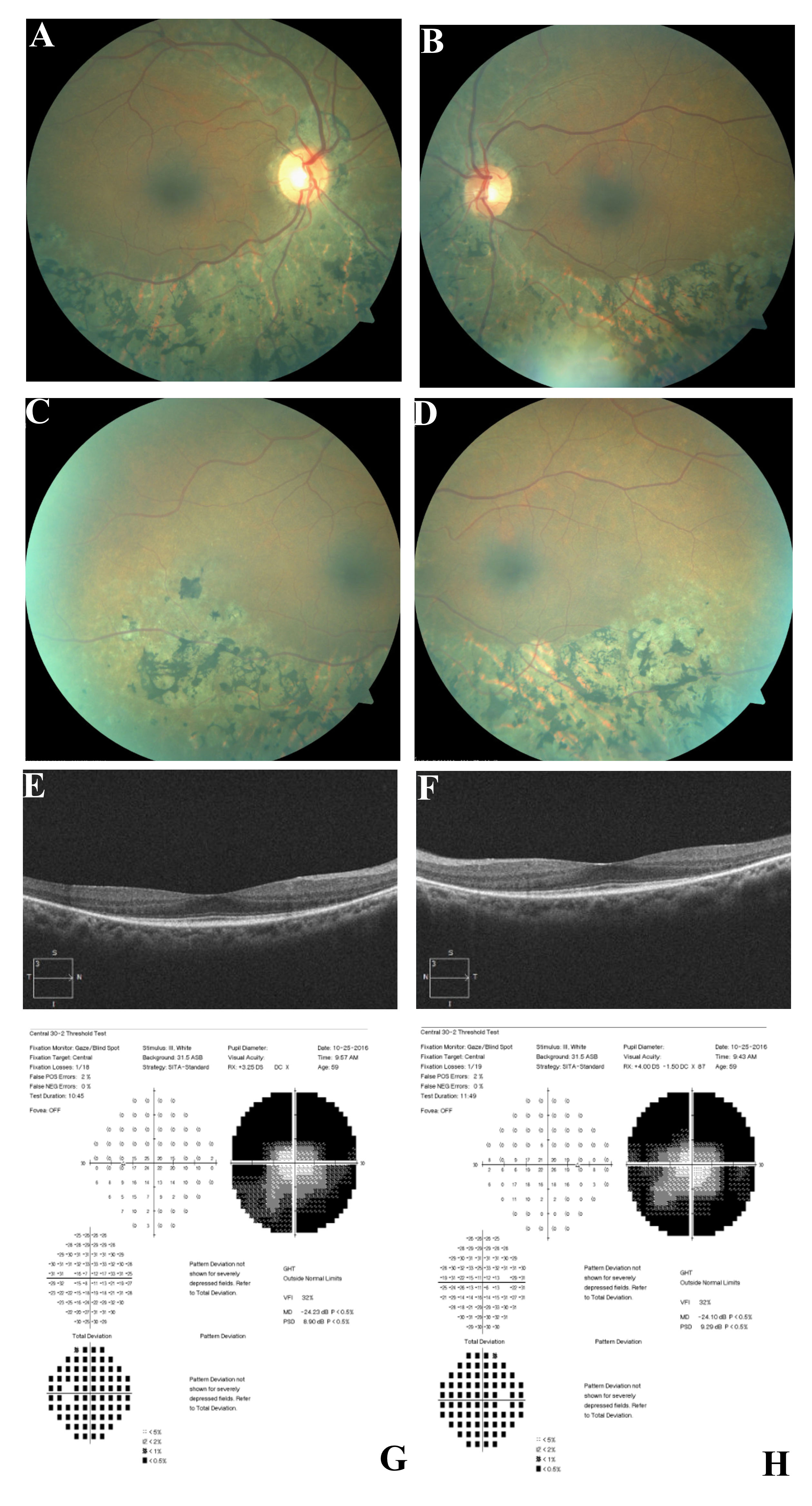
 Figure 6 of
Coussa, Mol Vis 2019; 25:869-889.
Figure 6 of
Coussa, Mol Vis 2019; 25:869-889.