Figure 5. Clinical imagining of patient 5 with a c.808A>C mutation in RHO (p.Ser270Arg). A, E: Oculus dexter (OD) color photos showing an oval-shaped island of RPE hypopigmentation and atrophy extending from within
the inferior posterior pole to the midperiphery and along the inferior arcades. B, F: Oculus sinister (OS) color photos showing an oval-shaped island of RPE hypopigmentation and atrophy extending from within
the inferior posterior pole to the midperiphery and along the inferior arcades. C, G: OD fundus autofluorescence (FAF) photos showing an oval-shaped island of RPE hypopigmentation and atrophy extending from
within the inferior posterior pole to the midperiphery and along the inferior arcades. D, H: OS FAF photos showing an oval-shaped island of RPE hypopigmentation and atrophy extending from within the inferior posterior
pole to the midperiphery and along the inferior arcades. I: OD foveal spectral-domain optical coherence tomography (SD-OCT) showing normal retinal structures. J: OS foveal SD-OCT showing normal retinal structures. K: OS Goldman visual field showing mild superior visual field loss. L: OD Goldman visual field showing mild superior visual field loss.
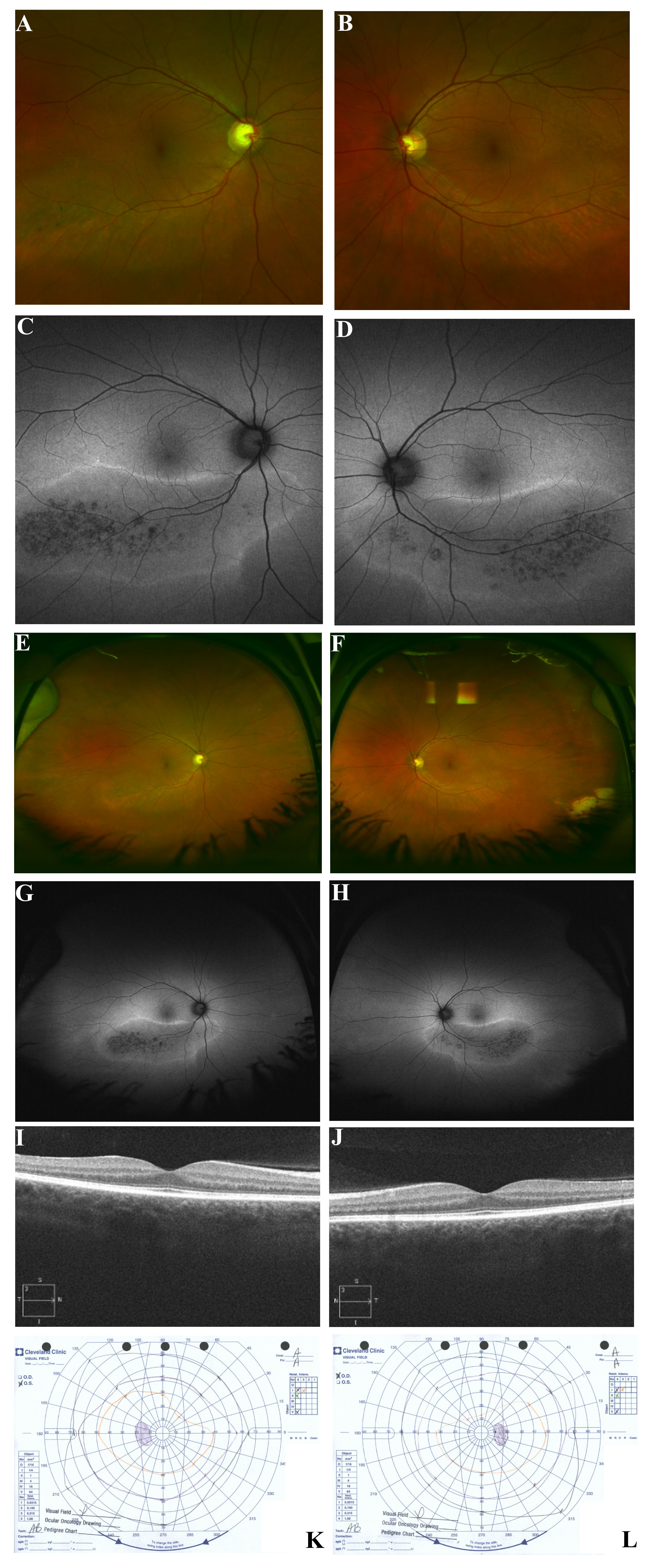
 Figure 5 of
Coussa, Mol Vis 2019; 25:869-889.
Figure 5 of
Coussa, Mol Vis 2019; 25:869-889.