Figure 4. Clinical imagining of patient 4 with a c.808A>C mutation in RHO (p.Ser270Arg). A, C: Ocular dexter (OD) color photo of posterior pole showing RPE hypopigmentation and atrophic changes with occasional bone
spicules most prominent along the inferior arcades. B, D: Oculus sinister (OS) color photo of posterior pole showing RPE hypopigmentation and atrophic changes with occasional bone
spicules most prominent along the inferior arcades. E, G: OD fundus autofluorescence (FAF) photos showing crescent-shaped hyper-autofluorescence (hyperAF) contouring the fovea inferiorly
and temporally as well as the optic nerve nasally. There is also a patchy pattern of hypoautofluorescence (hypoAF) along the
inferior arcades just adjacent to the previously mentioned crescent-shaped hyperAF. F, H: OS FAF photos showing crescent-shaped hyperAF contouring the fovea inferiorly and temporally as well as the optic nerve
nasally. There is also a patchy pattern of hypoAF along the inferior arcades just adjacent to the previously mentioned crescent-shaped
hyperAF. I: OD foveal SD-OCT showing parafoveal retinal thinning and loss of the ellipsoid zone and photoreceptor layer most prominent
in the inferior part of the posterior pole. J: OS foveal SD-OCT showing parafoveal retinal thinning and loss of the ellipsoid zone and photoreceptor layer most prominent
in the inferior part of the posterior pole. K: OS Goldman visual field showing superior parafoveal arcuate-like scotoma within the central 40 degrees. L: OD Goldman visual field showing superior parafoveal arcuate-like scotoma within the central 40 degrees.
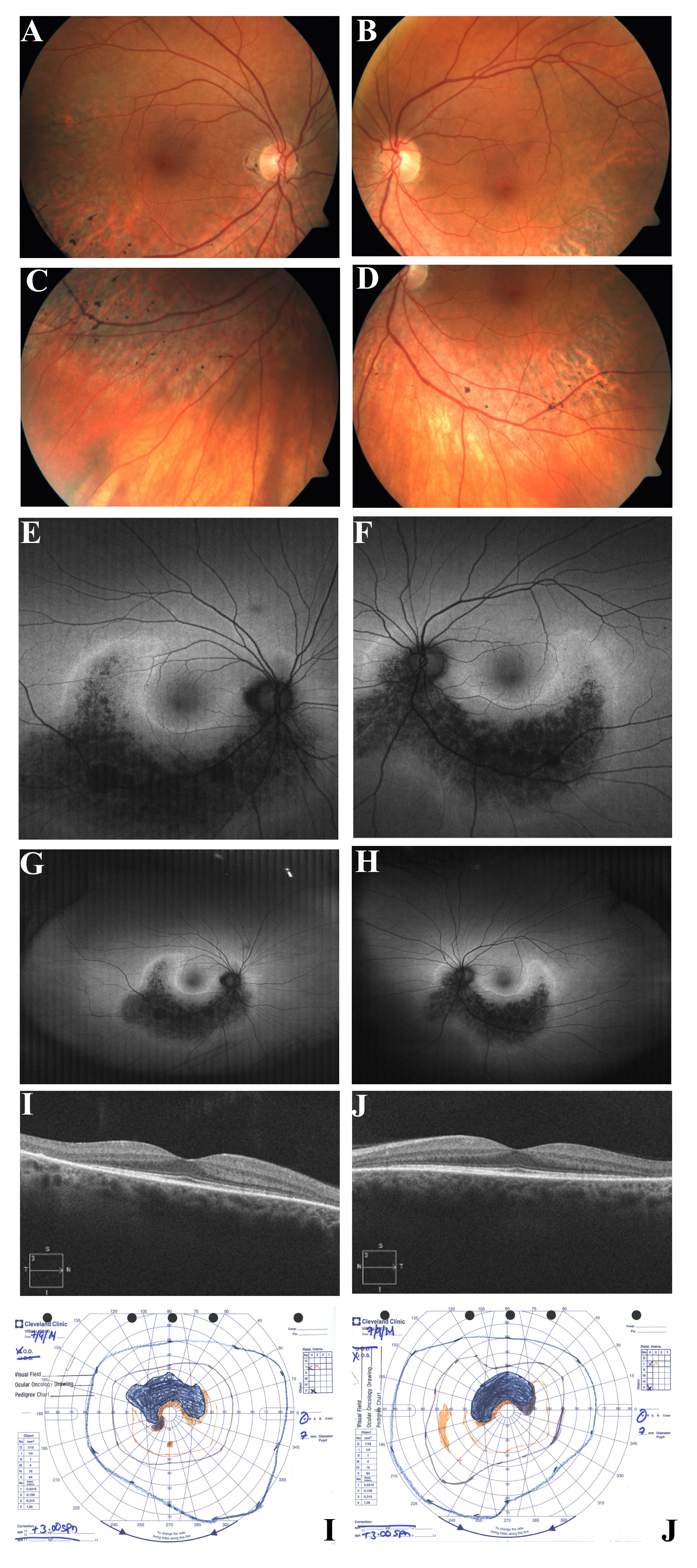
 Figure 4 of
Coussa, Mol Vis 2019; 25:869-889.
Figure 4 of
Coussa, Mol Vis 2019; 25:869-889.