Figure 10. Clinical imagining of patient 10 with a c.3092_3093delAG mutation in RPGR.A, C: Oculus dexter (OD) color photos showing a tessellated fundus, blunted foveal reflex, hypopigmentation, and atrophic changes
most prominent along the inferior arcades midperipherally. There are also marked bone spicules in the inferonasal midperiphery.
B, D: Oculus sinister (OS) color photos showing a tesselated fundus, blunted foveal reflex, hypopigmentation, and atrophic changes
most prominent along the inferior arcades midperipherally. There are also marked bone spicules in the inferonasal midperiphery.
E: OD fundus autofluorescence (FAF) photo showing a hypoautofluorescence (hypoAF) area bordering the inferior arcades and extending
inferiorly and inferonasally into the midperiphery. There is also a linear edge of hyper-autofluorescence (hyperAF) superior
to the area of hypoAF. F: OS FAF photo showing a hypoAF area bordering the inferior arcades and extending inferiorly and inferonasally into the midperiphery.
There is also a linear edge of hyperAF superior to the area of hypoAF. G: OD foveal spectral-domain optical coherence tomography (SD-OCT) showing generalized foveal thinning with marked ellipsoid
zone abnormalities as well as RPE hyperreflective round deposits and diffuse thickening of the RPE band. H: OS foveal SD-OCT showing generalized foveal thinning with marked ellipsoid zone abnormalities, as well as RPE hyperreflective
round deposits and diffuse thickening of the RPE band. I: OS Goldman visual field showing circumferential constriction with marked superior visual field defects. J: OD Goldman visual field showing circumferential constriction with marked superior visual field defects.
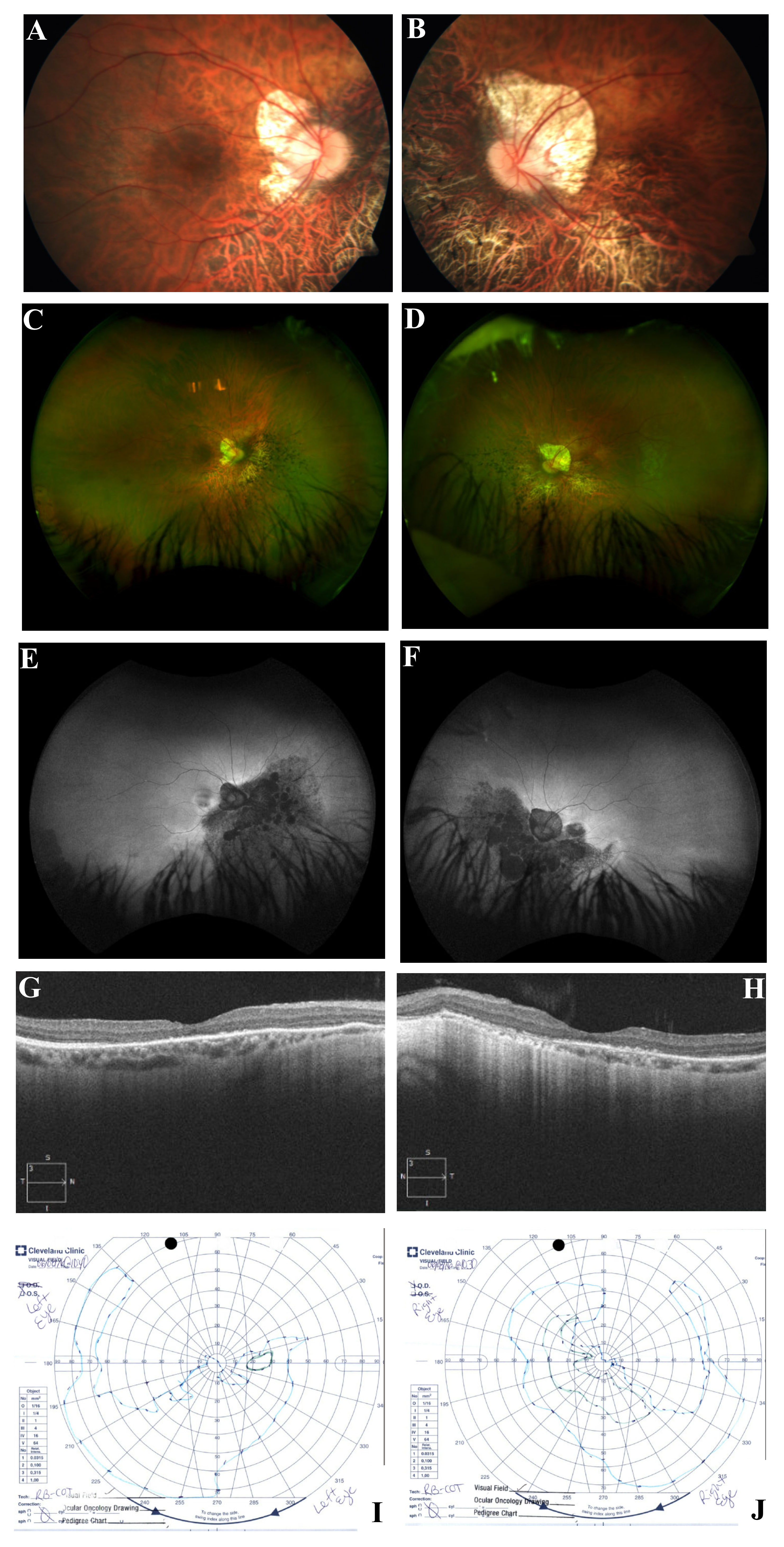
 Figure 10 of
Coussa, Mol Vis 2019; 25:869-889.
Figure 10 of
Coussa, Mol Vis 2019; 25:869-889.