Figure 1. Multimodal retinal imaging findings in TRPM1-associated CSNB in patient 1 with the novel homozygous missense c.1394T>A (p.Met465Lys) mutation. Top row: Color fundus of patient 1 right and left fundi. Middle row: Location of the optical coherence tomography line scan on the fundus en face image. Bottom row: Transfoveal single line optical coherence tomography scans of the right and left eyes of patient 1 demonstrating preserved
retinal lamination.
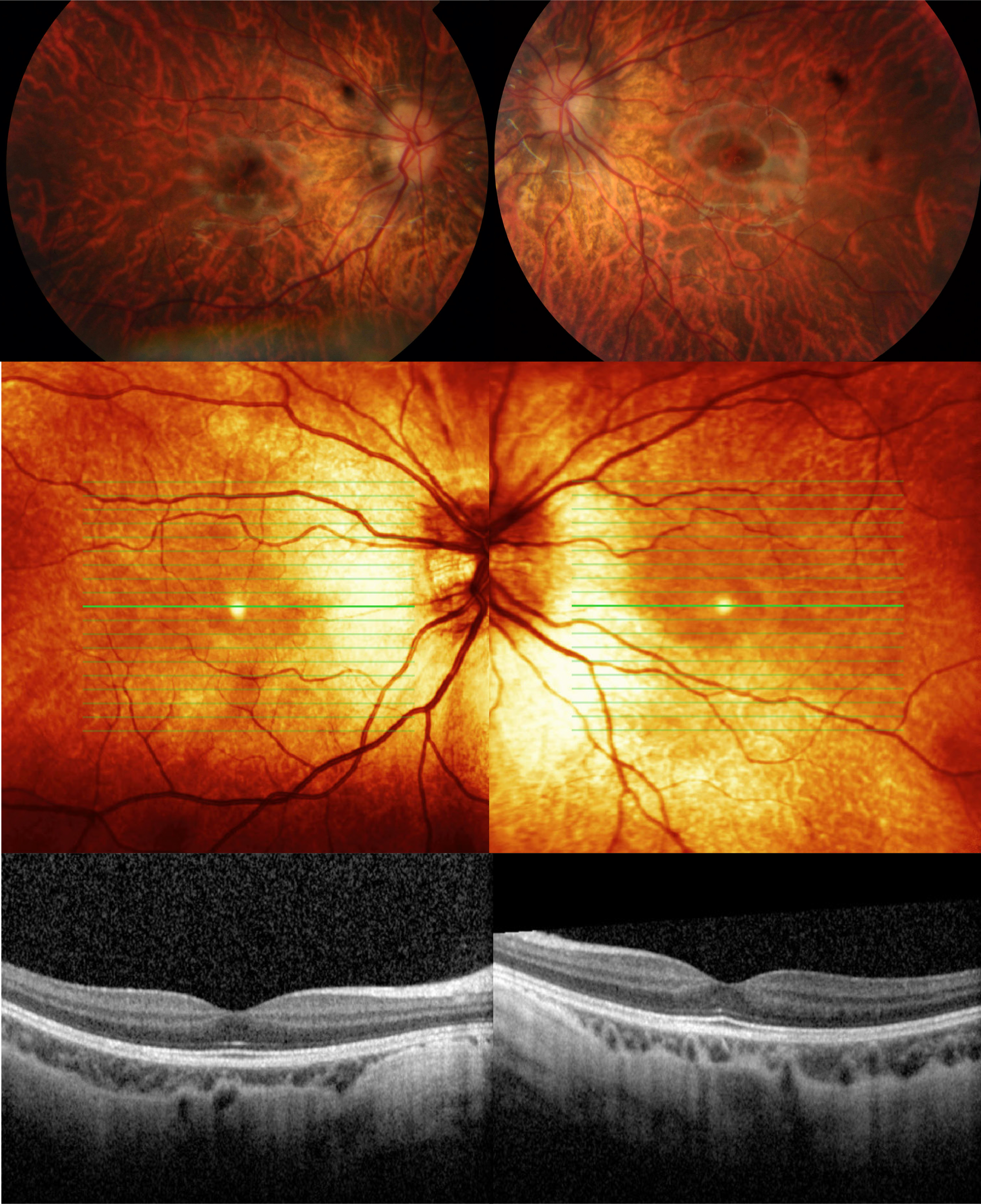
 Figure 1 of
Al-Hujaili, Mol Vis 2019; 25:851-858.
Figure 1 of
Al-Hujaili, Mol Vis 2019; 25:851-858.