Figure 3. Fundus images showing age-dependent changes in a patient (F07-II:1). The fundus appeared nearly normal in the posterior area,
with a few minor grayish-white spots in the midperiphery at the age of 3 years and 5 months (A), and progressed to significant yellowish-white spots in the midperiphery at the age of 7 years and 5 months (B, C). By the age of 8 years and 7 months, diffuse mottling hypopigmentation, with a few small intraretinal pigments, was visible
(D–I). Mild salt-and-pepper-like changes were observed (E, H).
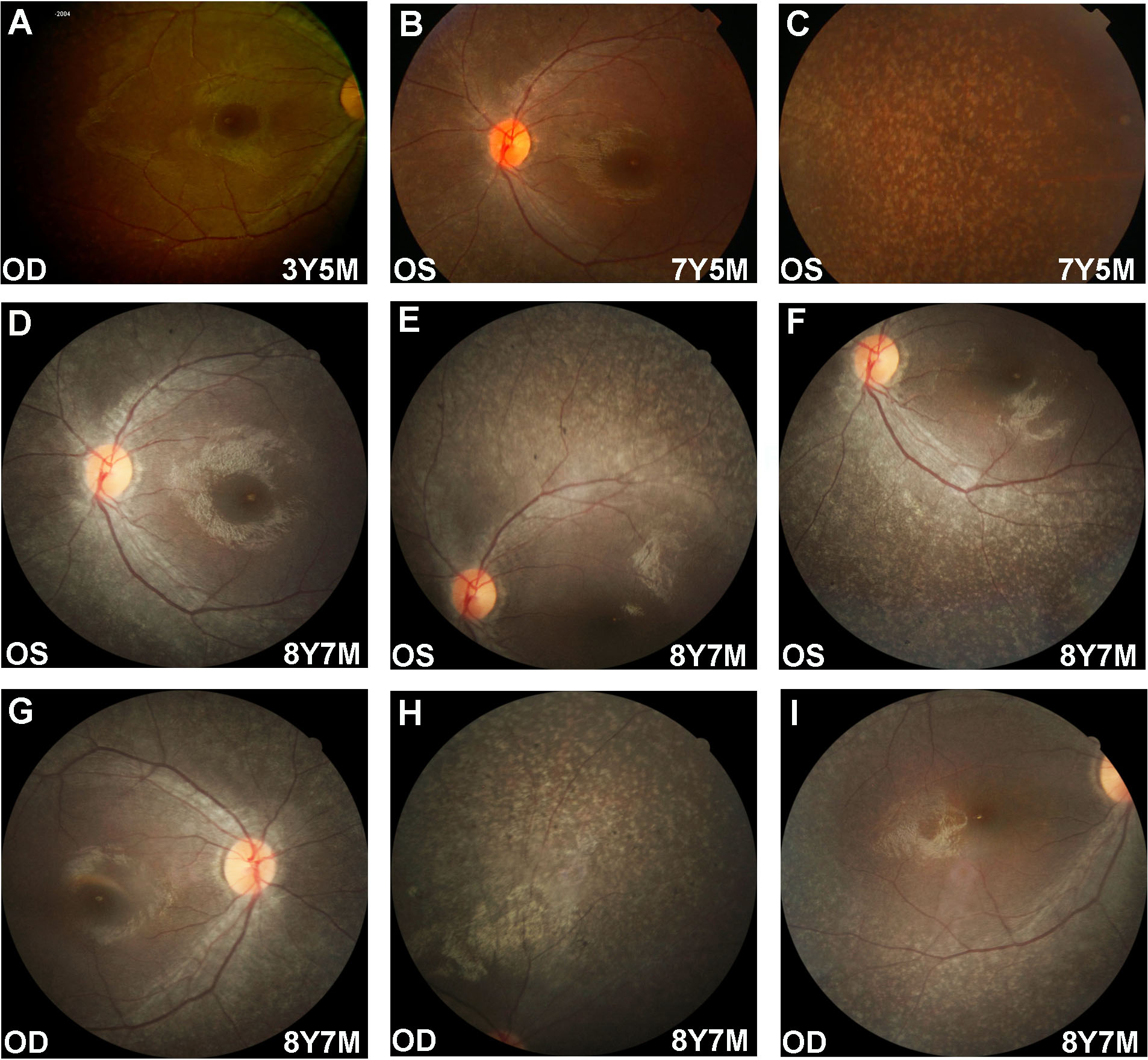
 Figure 3 of
Xiao, Mol Vis 2019; 25:821-833.
Figure 3 of
Xiao, Mol Vis 2019; 25:821-833.