Figure 2. Fundus images from patients with biallelic mutations in SPATA7. Representative fundus images show yellowish-white frosted degeneration in the midperiphery in patient F01-II:1 (A and B) and yellowish-white sand-like deposits in the midperiphery sparing the inferior area close to the optic fissure (C–F), in addition to yellowish-white mottled degeneration in the midperiphery (G–I). The homozygous novel mutation c.367C>T was identified in the F05-II:1, which is highlighted in red in G and H.
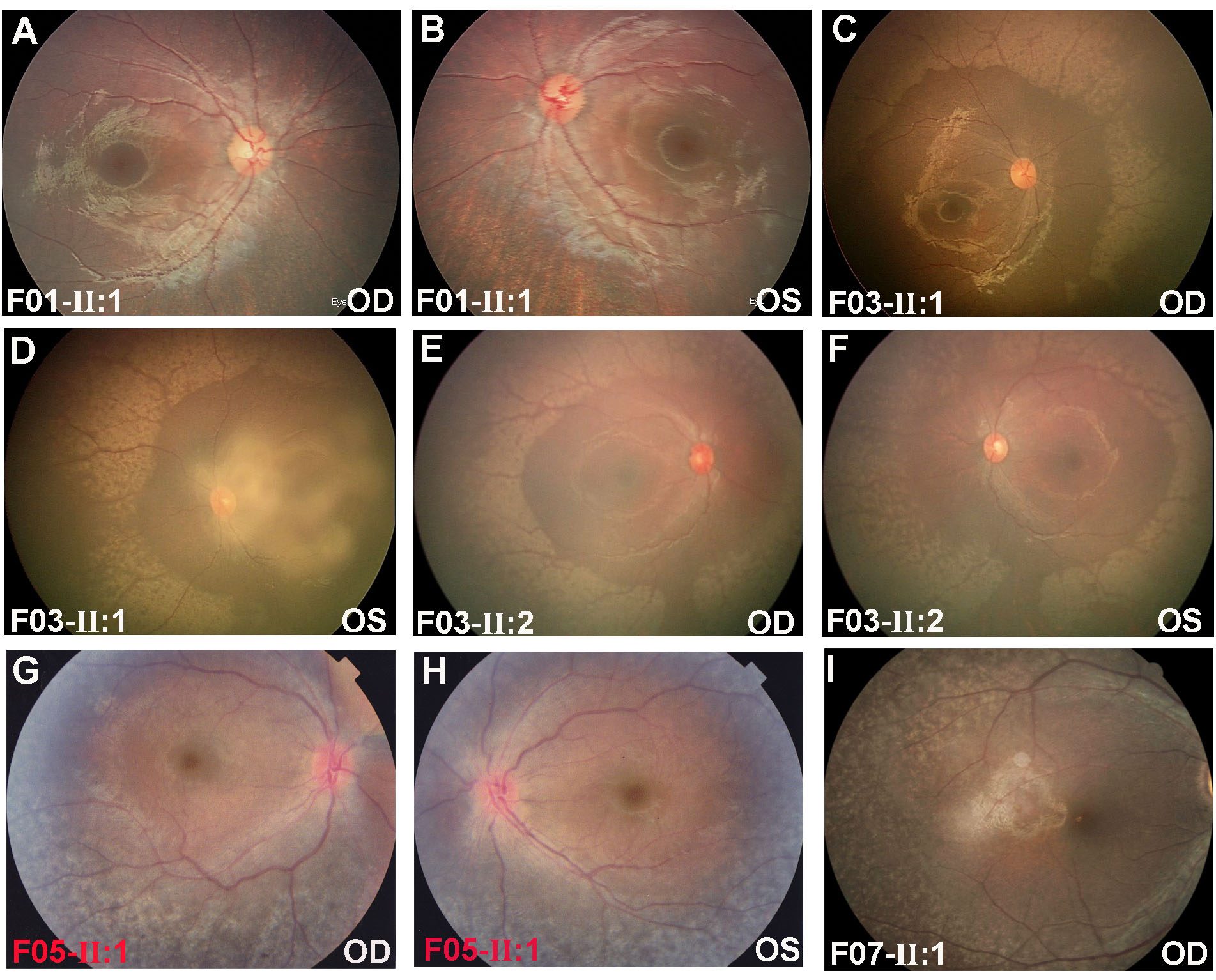
 Figure 2 of
Xiao, Mol Vis 2019; 25:821-833.
Figure 2 of
Xiao, Mol Vis 2019; 25:821-833.